CE/IVD antibodies for immunohistochemistry (IHC) in neuropathology are validated in vitro diagnostic reagents used to detect specific neuronal and glial antigens in formalin-fixed paraffin-embedded (FFPE) tissue. Peer-reviewed neuropathology literature supports their role in improving reproducibility and diagnostic accuracy in central nervous system (CNS) diseases, including brain tumors and neurodegenerative disorders.
Biological Significance
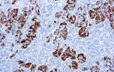
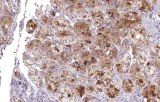
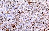
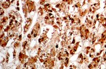
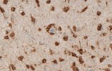
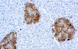
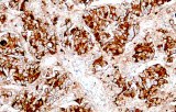
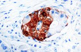
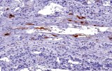
Targeted proteins such as GFAP, OLIG2, NeuN, synaptophysin, neurofilament, and Iba1 reflect astrocytic, oligodendroglial, neuronal, and microglial lineages. Their expression patterns are widely used in peer-reviewed studies to define CNS cell identity, differentiation state, and neuroinflammatory or degenerative processes.
Diagnostic Utility in Neuropathology
CE/IVD IHC antibodies support CNS tumor classification (gliomas, embryonal tumors, metastases), assist in differential diagnosis, and help identify proteinopathies associated with Alzheimer’s disease and other neurodegenerative conditions. They are routinely used in biomarker panels for tumor grading and diagnostic stratification in neuro-oncology.
Key Features of CE/IVD Antibodies
These reagents are manufactured and validated under regulatory in vitro diagnostic standards to ensure specificity, sensitivity, and lot-to-lot consistency.
- Optimized for formalin-fixed paraffin-embedded (FFPE) tissue samples.
- Compatible with manual and automated immunohistochemistry (IHC) platforms.
- Designed for standardized clinical diagnostic workflows.
- Validated for reproducible antigen detection in neuropathology applications.