

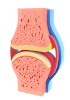
Human synovium tissue pieces and blocks are solid, three-dimensional specimens of the synovial membrane harvested from human donors and supplied for research and development applications. The synovium, or synovial membrane, is a specialized connective tissue that lines the inner surface of diarthrodial joints and represents the deep surface of the joint capsule. It is composed of areolar connective tissue containing elastic fibres and functions to secrete synovial fluid, which lubricates and nourishes the avascular articular cartilage. Beyond its mechanical role, the synovium serves as a critical immunological barrier that defends against infection and participates in inflammatory responses. In many synovial joints, the membrane accumulates adipose tissue in structures known as adipose bodies, the infrapatellar fat body of the knee being a prominent example. The resident cells of the synovium, termed synoviocytes, play a primordial role in the function of the hemosynovial barrier and are thought to be the first to phagocytose crystals and trigger the inflammatory process. Inflammation of the synovial membrane, known as synovitis, is a central feature of arthropathies including osteoarthritis (OA), rheumatoid arthritis (RA), septic arthritis, lupus erythematosus, and gout.
Histological Organization and Cellular Composition
The synovium exhibits a distinctive histological architecture that underlies its functional specialization. The normal synovial lining layer (intima) is one to three cells thick and is composed of closely associated macrophage-like (type A) and fibroblast-like synoviocytes (type B) that are not separated from the underlying stroma by a basement membrane. The intimal lining cells express distinctive markers, including CD68 on macrophages and CD55 on fibroblast-like synoviocytes. The subintimal tissue (sublining layer) is separated from the intima by an intermediate fibrillar zone rich in type III collagen, while basement membrane-like material associated with synovial lining cells stains for type IV collagen and laminin. Depending on the stromal and cellular content of the sublining layer, the synovial tissue can be classified as areolar, adipose, or fibrous. In aged synovium, three main lining cell phenotypes can be recognized: synthetic type (present at all ages and hypertrophied in aged subjects), macrophage-like (increasing with age), and fibroblast-like, with collagen content increasing with age.