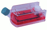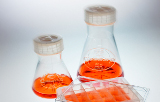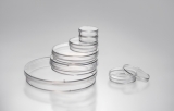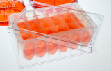

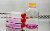
Cell culture plastics constitute the physical interface between in vitro biological systems and their experimental environment. While numerous polymers have been employed historically—including polyethylene terephthalate (PET), polyethylene (PE), polyvinyl chloride (PVC), and polypropylene (PP)—polystyrene (PS) remains the predominant material due to its low cost, optical clarity, and chemically inert nature. However, the biological performance of PS is not intrinsic; it is profoundly modified by surface engineering and fabrication parameters that dictate cell attachment, proliferation, and phenotypic maintenance.
Polymer Selection and Optical Properties
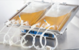
The choice of base polymer is governed by application-specific requirements. Polystyrene offers excellent optical transparency, making it indispensable for phase-contrast and fluorescence microscopy. Its heat distortion point of 64–80°C renders it unsuitable for autoclaving but compatible with gamma irradiation and gas plasma sterilization. Polycarbonate, by contrast, withstands autoclaving (heat distortion 138–143°C) but exhibits lower optical clarity, whereas polypropylene provides superior chemical resistance for applications involving organic solvents. High-density polyethylene possesses high gas permeability, a property exploited in oxygen-permeable culture bags and membranes, while polystyrene exhibits low oxygen permeability (coefficient approximately 2.6 × 10⁻¹¹ mL(O2)·cm/(cm2·sec·mmHg)).
Surface Treatment Technologies
Native polystyrene is hydrophobic, with a surface energy of approximately 20–40 mN/m, which resists the adsorption of adhesion-mediating proteins and prevents anchorage-dependent cells from attaching. Tissue culture (TC) treatment addresses this limitation through two principal methods: corona discharge (employed for flasks, dishes, and microplates) and gas plasma treatment (used for roller bottles and culture tubes). Both processes generate highly energetic oxygen ions that graft onto the polystyrene surface, cleaving polymer chains and introducing oxygen-containing functional groups—specifically hydroxyl (–OH) and carboxyl (–COOH) moieties—as well as nitrogen-containing amine groups. The resulting surface becomes hydrophilic and negatively charged when immersed in culture medium, enabling the adsorption of serum-derived proteins such as fibronectin and vitronectin, which in turn mediate integrin-dependent cell adhesion.