Anti-p120 catenin (p120-ctn) immunohistochemistry (IHC) is an established ancillary diagnostic tool used in surgical pathology to evaluate cell–cell adhesion dynamics and to assist in lineage and subtype classification of epithelial neoplasms, most notably in distinguishing invasive lobular carcinoma (ILC) from invasive ductal carcinoma (IDC) of the breast.
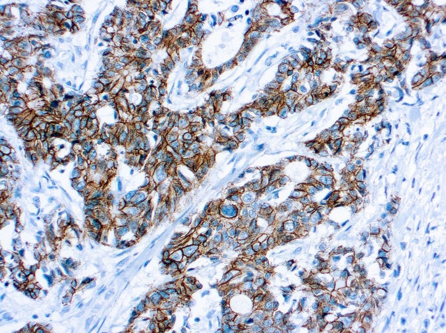
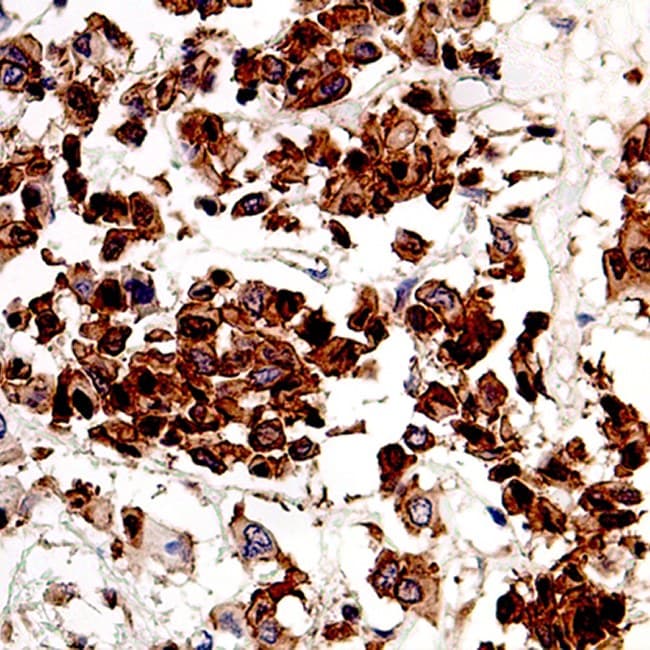
p120 is a cadherin-associated protein that stabilizes E-cadherin at adherens junctions and, when mislocalized, provides a robust IHC signal pattern (membranous vs cytoplasmic) that is diagnostically meaningful.
Anti-p120 reagents provided as CE-marked / IVD-intended kits deliver standardized, validated reagents for use on FFPE tissue in clinical laboratories.
Clinical utility In breast pathology
- Differentiation of lobular vs ductal neoplasia: p120 localization is a powerful adjunct: membranous p120 staining is typical of ductal phenotype, whereas cytoplasmic and loss of membranous p120 is characteristic of lobular neoplasia (including LCIS and ILC), even in cases with ambiguous or aberrant E-cadherin staining. This makes p120 especially useful for early/atypical lesions and metastatic workups where morphology alone is equivocal.
- Complementary marker: p120 is commonly used in a diagnostic panel together with E-cadherin (CDH1) and β-catenin to increase sensitivity and specificity for ILC vs IDC. Use of the panel reduces false-negative/false-positive calls associated with single-antibody interpretation.
Clinical utility In gynecological pathology
- Assessment of cadherin–catenin complex alterations: altered expression/localization of p120 has been documented in ovarian and endometrial carcinomas and has been correlated with cadherin switching and tumor progression in subsets of gynecologic malignancies. While not a routine standalone diagnostic for most gynecologic tumors, p120 IHC can contribute to:
• subclassification when epithelial differentiation is uncertain.
• mechanistic research panels for epithelial–mesenchymal transition (EMT) and cadherin switching in ovarian carcinoma.
• resolving discordant E-cadherin staining patterns.
Key features of Anti-p120 Catenin CE/IVD antibodies
- CE-marked / IVD intended: supplied and validated for diagnostic IHC on FFPE tissues.
- Distinctive staining patterns: reliable membranous staining in cadherin-intact epithelia versus cytoplasmic redistribution in lobular phenotype, high diagnostic value for lobular vs ductal determination.
- Compatibility with E-cadherin/β-catenin panels: validated to be used side-by-side with E-cadherin and other adhesion markers for increased diagnostic accuracy.
- Clones and formats: multiple well-characterized clones are available (research and IVD formats); consult the specific product datasheet for clone information, recommended antigen retrieval, positive/negative controls, and dilution/pretreatment instructions.
- Applications: FFPE surgical biopsy and resection specimens; primary tumor typing and metastatic site workups; research into cadherin-catenin biology and EMT.
Why include Anti-p120 on your diagnostic panel?
- Enhances accuracy in diagnosing invasive lobular carcinoma (ILC).
- Helps resolve discordant E-cadherin staining.
- Supports research and biomarker studies of cadherin-catenin dysregulation in breast and gynecologic cancers.