Glypican-3 (GPC3) is a cell-surface heparan sulfate proteoglycan that is anchored to the plasma membrane through a glycosylphosphatidylinositol (GPI) linkage. It belongs to the glypican family of proteoglycans, which play key roles in regulating cell–cell and cell–matrix interactions during development and disease.
Biological Significance of Glypican-3
Biologically, GPC3 modulates signaling pathways involved in cell proliferation, differentiation, migration, and apoptosis. Through its heparan sulfate side chains, GPC3 can influence multiple growth-regulatory pathways, most notably Wnt/β-catenin signaling, by facilitating ligand–receptor interactions at the cell surface. Additional interactions with growth factor–related signaling pathways have been described, although the precise mechanisms remain incompletely defined.
GPC3 is considered an oncofetal protein. It is highly expressed during embryonic development, particularly in fetal liver, but is largely absent or present at very low levels in most normal adult tissues. Re-expression of GPC3 in malignant tissues reflects dysregulation of developmental signaling programs during tumorigenesis.
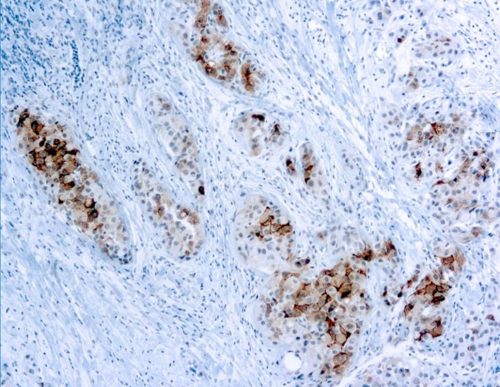
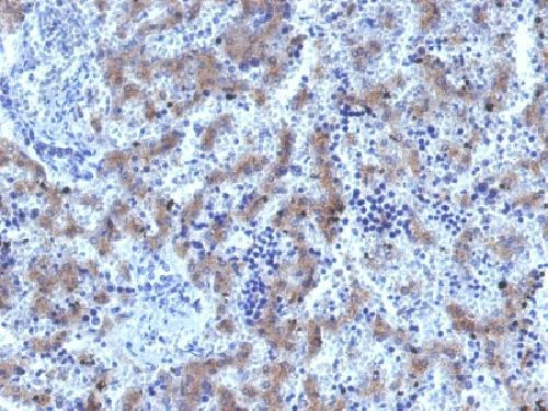
In hepatic pathology, GPC3 is frequently overexpressed in hepatocellular carcinoma (HCC) compared with non-neoplastic liver tissue. Immunohistochemical studies consistently demonstrate cytoplasmic and/or membranous GPC3 staining in a substantial proportion of HCCs, whereas normal adult hepatocytes are typically negative.
Expression of GPC3 in benign hepatocellular lesions, such as regenerative or cirrhotic nodules, is uncommon but not entirely absent. Occasional weak or focal staining has been reported, particularly in dysplastic nodules, indicating that GPC3 expression is highly suggestive of malignancy but not absolutely specific for HCC.
Diagnostic Utility in Gastrointestinal Pathology
In gastrointestinal and hepatic pathology, GPC3 immunohistochemistry (IHC) is a well-established ancillary tool, particularly for the diagnosis of hepatocellular carcinoma. Reported diagnostic sensitivities vary across studies and specimen types, while specificity is generally high when differentiating HCC from benign hepatic lesions and most metastatic tumors to the liver.
Importantly, negative GPC3 staining does not exclude HCC, especially in well-differentiated tumors or limited biopsy material. Consequently, GPC3 is most effective when used as part of a multimarker immunohistochemical panel, commonly in combination with markers of hepatocellular differentiation and malignant transformation.
Beyond the liver, GPC3 expression has been documented in a minority of non-hepatic neoplasms, including certain gastrointestinal and pancreatic epithelial tumors as well as tumors with hepatoid differentiation. Although uncommon, such expression represents a potential diagnostic pitfall, particularly when evaluating metastatic lesions involving the liver.
Key Features of Anti-Glypican-3 CE/IVD Antibodies
Anti-GPC3 antibodies developed for CE-marked and in vitro diagnostic (IVD) use are optimized for detection of GPC3 protein in formalin-fixed, paraffin-embedded (FFPE) tissue sections, which are standard in routine diagnostic pathology.
Clinically validated anti-GPC3 antibodies typically demonstrate membranous and/or cytoplasmic staining patterns consistent with known GPC3 localization. Performance characteristics, including sensitivity and staining intensity, may vary depending on the antibody clone, antigen-retrieval conditions, and staining platform.
Key features of CE/IVD-validated anti-GPC3 antibodies include:
- High analytical specificity for GPC3 protein when appropriate controls are applied
- Reproducible staining performance suitable for routine diagnostic workflows
- Compatibility with automated immunostaining systems, supporting standardization and inter-laboratory consistency
When incorporated into comprehensive immunohistochemical panels, anti-GPC3 antibodies contribute meaningfully to the differential diagnosis of hepatocellular carcinoma and selected gastrointestinal and hepatobiliary lesions, improving diagnostic confidence in routine clinical practice.