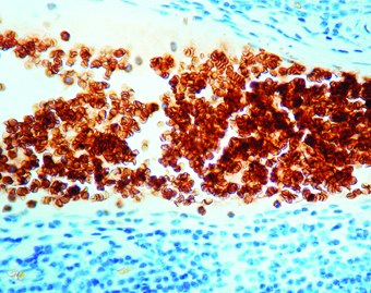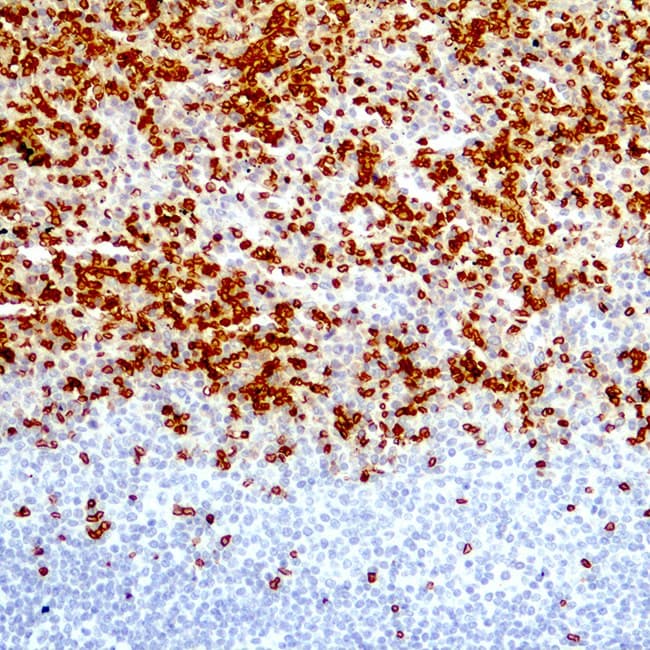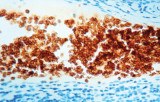
Anti-Glycophorin A (CD235a) CE/IVD antibodies are widely used immunohistochemistry (IHC) reagents in hematopathology for the identification of erythroid lineage cells in bone marrow and extramedullary tissues. Glycophorin A (GPA) is the major sialoglycoprotein of the erythrocyte membrane, characterized by highly restricted expression within the erythroid lineage across most stages of maturation, making it a reliable and widely used biomarker in diagnostic pathology.
Biological Significance of Glycophorin A
Glycophorin A plays a central role in erythroid cell biology and membrane structure:
- Erythroid-associated membrane protein expressed on erythrocytes and their precursors, with highly limited expression outside the erythroid lineage.
- Carries MN blood group antigens and contributes to red cell membrane stability and negative surface charge through sialic acid residues.
- Expressed during erythroid differentiation, with increasing intensity from early erythroblasts to mature erythrocytes.
- Serves as a reliable indicator of erythropoiesis in bone marrow and peripheral tissues when interpreted in conjunction with morphology and additional markers.
Diagnostic Utility in Hematopathology
Anti-Glycophorin A IHC antibodies are established tools for identifying erythroid lineage in both reactive and neoplastic conditions:
- Detection of erythroid precursors in bone marrow biopsies, supporting lineage assignment in hematologic disorders.
- Identification of erythroid differentiation in acute leukemias, typically used alongside complementary erythroid markers (e.g., hemoglobin, CD71).
- Evaluation of myelodysplastic syndromes (MDS) through assessment of the erythroid compartment and maturation patterns.
- Differentiation of erythroid cells from myeloid, lymphoid, and megakaryocytic populations in diagnostically challenging cases.
- Recognition of late-stage erythroblasts and mature erythrocytes, typically showing membranous staining in FFPE tissues.
Key Features of Glycophorin A CE/IVD Antibodies
- High specificity for erythroid lineage when used under optimized and validated conditions.
- Suitable for formalin-fixed, paraffin-embedded (FFPE) tissues within routine diagnostic workflows.
- Typically produce membranous staining consistent with erythrocyte membrane localization.
- Validated on major automated IHC platforms, depending on manufacturer instructions for use (IFU).
- Available as CE/IVD-labeled reagents, supporting standardized and reproducible application in accredited hematopathology laboratories.