Anti-Fascin CE/IVD antibodies are commonly used immunohistochemical (IHC) reagents for the evaluation of dendritic cell differentiation and related neoplasms in hematopathology. Fascin (FSCN1) is a ~55 kDa actin-bundling protein with relatively restricted expression in hematolymphoid tissues, predominantly in dendritic cells, supporting its utility as a lineage-associated marker when interpreted in context.
Biological Significance of Fascin
Fascin plays a key role in cytoskeletal organization and immune cell function:
- Actin-bundling protein that organizes filamentous actin into parallel arrays, facilitating filopodia formation and cell motility.
- Highly expressed in mature dendritic cells, with minimal expression in most resting lymphocytes.
- Contributes to dendritic cell maturation, migration, and antigen-presenting function.
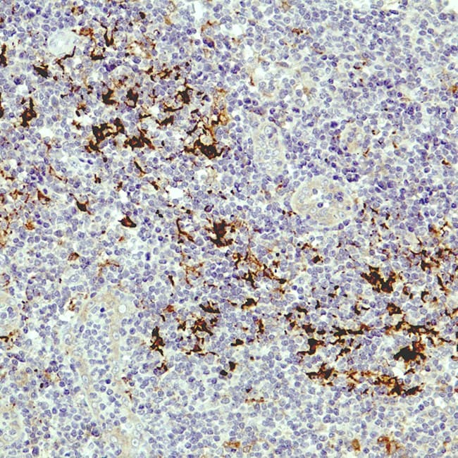
- Localized in interdigitating dendritic cells within lymphoid tissues.
- Also expressed in a range of non-hematopoietic cells and upregulated in multiple malignancies, where it is associated with increased cellular motility and invasiveness.
Diagnostic Utility of Fascin in Hematopathology
Fascin IHC serves as an ancillary marker for dendritic lineage and selected lymphoid neoplasms:
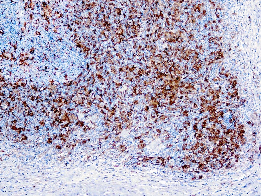
- Strong cytoplasmic expression in classical Hodgkin lymphoma (Reed–Sternberg cells).
- Supports distinction between classical Hodgkin lymphoma and nodular lymphocyte-predominant Hodgkin lymphoma when used as part of a comprehensive immunophenotypic panel.
- Highlights dendritic cell populations and meshworks in lymphoid tissues and can assist in identifying dendritic cell neoplasms.
- Commonly used in combination with markers such as CD1a, S100, CD21, and CD23 for lineage assessment.
- Aids architectural evaluation in reactive and neoplastic lymphoid conditions.
Key Features of Anti-Fascin CE/IVD Antibodies
- Optimized for formalin-fixed, paraffin-embedded (FFPE) tissue sections.
- Produce distinct cytoplasmic staining in fascin-expressing cells.
- Sensitive detection of fascin expression, requiring interpretation within a panel due to limited specificity.
- Compatible with standard antigen retrieval and detection systems.
- Require appropriate positive and negative controls (e.g., dendritic cells in lymphoid tissue).
- Intended for in vitro diagnostic (IVD) use with expert pathological interpretation.