Epithelial membrane antigen (EMA), corresponding to MUC1, is a high-molecular-weight, heavily glycosylated transmembrane mucin expressed on the apical surface of many epithelial cells. Its structural and functional properties have been extensively characterized in peer-reviewed literature, supporting roles in cell adhesion, intracellular signaling, and epithelial organization.
Biological Significance of EMA (MUC1)
- EMA/MUC1 is a type I transmembrane glycoprotein characterized by extensive O-linked glycosylation, which contributes to its antigenic and structural properties.
- It participates in modulation of cell–cell and cell–matrix interactions, as well as intracellular signaling pathways.
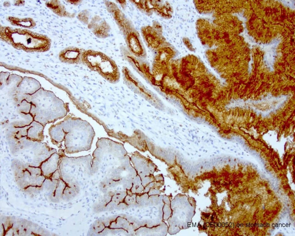
- In normal epithelial tissues, MUC1 is typically apically polarized, whereas in malignant transformation it often shows loss of polarity, overexpression, and altered glycosylation.
- Aberrant glycosylation patterns are associated with tumor-associated phenotypes and changes in cellular behavior.
Diagnostic Utility in Hematopathology
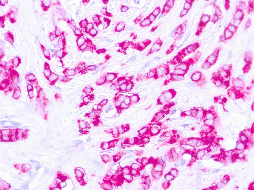
- EMA is commonly used in immunohistochemistry (IHC) panels as a marker to support identification of epithelial differentiation in the evaluation of neoplasms.
- It can assist in distinguishing carcinomas from many hematolymphoid and mesenchymal tumors, but it is not entirely specific.
- EMA expression may be observed in certain hematopoietic neoplasms, including plasma cell neoplasms (e.g., plasmacytoma) and in subsets of lymphomas, where it can support diagnostic interpretation.
- In practice, EMA is interpreted alongside other markers in a broader panel, particularly in cases of poorly differentiated malignancies, to help determine lineage when morphology is inconclusive.
Key Features of EMA CE/IVD Antibodies
- Intended for in vitro diagnostic (IVD) use when appropriately validated within regulated laboratory workflows; CE marking indicates conformity with applicable regulatory requirements in relevant jurisdictions (product- and manufacturer-dependent).
- Optimized for detection in formalin-fixed, paraffin-embedded (FFPE) tissues, supporting routine histopathology workflows.
- Designed to produce membranous and/or cytoplasmic staining patterns, consistent with the known subcellular localization of MUC1/EMA in tissue sections.
- Performance characteristics such as sensitivity, specificity, and reproducibility depend on validated protocols, including antigen retrieval conditions, antibody clone, detection system, and laboratory quality controls.