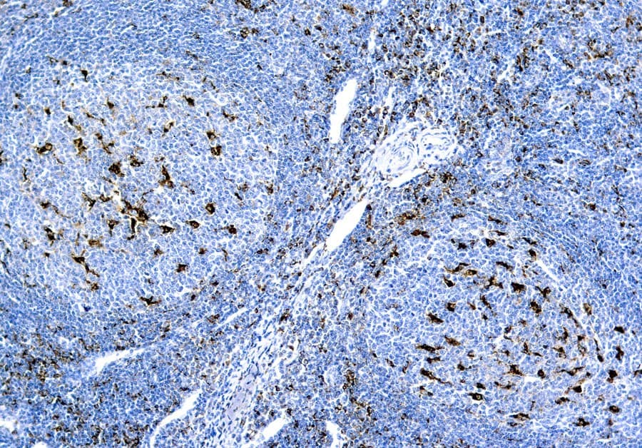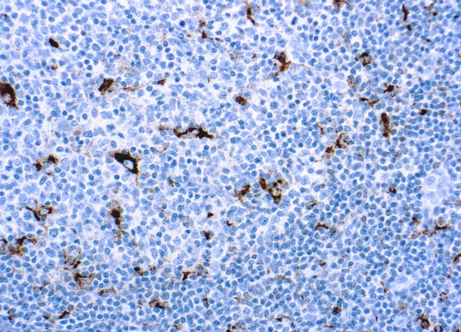
Cluster of Differentiation 68 (CD68) is a heavily glycosylated transmembrane glycoprotein predominantly expressed in cells of the monocyte–macrophage lineage. CE/IVD validated CD68 antibodies for immunohistochemistry (IHC) enable reliable identification and quantification of macrophages and histiocytic populations in formalin-fixed, paraffin-embedded (FFPE) tissue, supporting hematopathology diagnostics, disease classification, and research into immune microenvironments.
Biological Significance of CD68
- Mononuclear phagocyte marker: Highly expressed in macrophages, Kupffer cells, Langerhans cells, and osteoclasts.
- Lysosomal association: Member of the lysosome-associated membrane glycoprotein (LAMP) family, localized primarily to lysosomes and endosomes, reflecting phagocytic function.
- Expression nuances: May be detected in some non-macrophage cells (e.g., dendritic cells, fibroblasts) depending on tissue context and antibody clone. Plasma membrane expression can occur in activated macrophages.
Diagnostic Utility
- Macrophage identification: Highlights macrophages in bone marrow, lymphoid tissue, and histiocytic lesions, aiding differentiation from lymphoid or non-hematopoietic neoplasms.
- Inflammation and microenvironment analysis: Supports quantification of macrophage infiltrates, including tumor-associated macrophages (TAMs), providing prognostic and research insights.
- Hematologic neoplasms: Positive CD68 staining contributes to diagnosis of histiocytic sarcoma, myelomonocytic leukemia, and related disorders.
Key Features of CD68 CE/IVD Antibodies
- CE/IVD validated: Optimized for robust detection in FFPE tissue with reproducible cytoplasmic staining.
- Clone selection: Monoclonal clones (e.g., KP1, PGM1) offer high specificity; clone choice influences tissue reactivity and staining pattern.
- Diagnostic integration: Best interpreted alongside complementary markers and morphology for accurate hematopathology evaluation.