Cluster of differentiation 3 (CD3) is a key component of the T-cell receptor (TCR) signaling complex and a widely used pan-T-cell marker in diagnostic hematopathology. In immunohistochemistry (IHC), anti-CD3 antibodies enable reliable identification of T-cell populations in formalin-fixed paraffin-embedded (FFPE) tissues and play an important role in the classification of lymphoid malignancies and the evaluation of immune infiltrates.
Biological Significance of CD3
CD3 complex is a multimeric protein complex associated with the T-cell receptor and composed of invariant chains (γ, δ, ε, and ζ) that mediate intracellular signaling following antigen recognition. These chains contain immunoreceptor tyrosine-based activation motifs that initiate downstream signaling pathways after TCR engagement.
Key biological roles include:
- T-cell activation and signal transduction: CD3 transmits activation signals from the TCR to intracellular signaling pathways, enabling T-cell activation, proliferation, and effector functions.
- T-cell lineage identification: CD3 is expressed on nearly all mature T lymphocytes and during multiple stages of T-cell development, making it a reliable marker of T-cell lineage.
- Immune response regulation: The TCR–CD3 complex is essential for antigen recognition and the initiation of adaptive immune responses.
Diagnostic Utility of CD3 in Hematopathology
In hematopathology, CD3 immunostaining is widely used to identify and characterize T-cell populations in tissue samples.
Major diagnostic applications include:
- Determination of lymphoid lineage: CD3 positivity supports confirmation of T-cell lineage, helping distinguish T-cell lymphomas and leukemias from B-cell or myeloid malignancies.
- Characterization of lymphoproliferative disorders: CD3 staining highlights T-cell populations within lymphoid tissues and bone marrow.
- Evaluation of T-cell infiltrates: CD3 IHC enables visualization and assessment of T-cell infiltration in tissues and tumor microenvironments.
Key Features of Anti-CD3 CE/IVD Antibodies for IHC
- Optimized for FFPE tissue sections, the standard specimen type in diagnostic pathology.
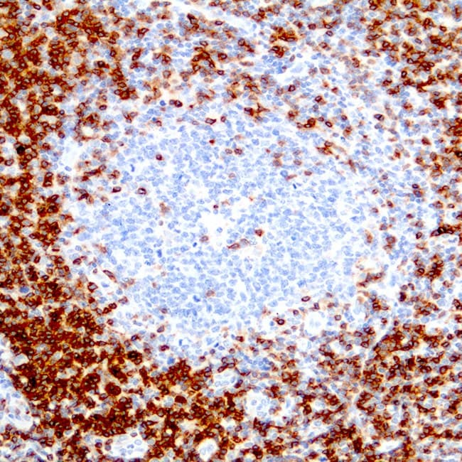
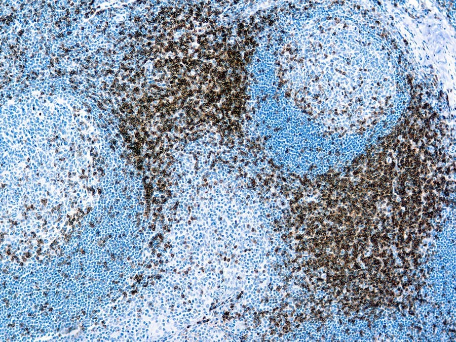
- Specific detection of CD3-expressing T lymphocytes, typically producing membranous staining in mature T cells, with cytoplasmic staining possible depending on antibody clone and cellular context.
- High sensitivity for pan-T-cell identification in lymphoid tissues.
- Compatibility with routine IHC workflows and use in diagnostic panels with markers such as CD20 and CD45.