Anti-CD15 antibodies are widely used immunohistochemical reagents for detecting CD15 (Lewis X antigen) in hematopathology. CD15 immunostaining is routinely applied to evaluate granulocytic differentiation and Hodgkin lymphoma–associated tumor cells in formalin-fixed, paraffin-embedded (FFPE) tissue sections. When incorporated into multiparametric immunohistochemical panels, CD15 contributes to the immunophenotypic characterization and differential diagnosis of hematologic malignancies. Reliable detection of CD15 is therefore an important component of diagnostic workflows in pathology laboratories.
Biological Significance of CD15
CD15, also known as Lewis X (3-fucosyl-N-acetyllactosamine), is a carbohydrate antigen expressed on glycoproteins and glycolipids located on the surface of leukocytes.
Key biological characteristics include:
- High expression on mature granulocytes, particularly neutrophils and eosinophils, with lower expression on monocytes.
- Regulation through glycosylation pathways, including the activity of α1,3-fucosyltransferases involved in myeloid cell differentiation.
- Participation in leukocyte adhesion and trafficking, contributing to inflammatory and immune responses through interactions with selectins.
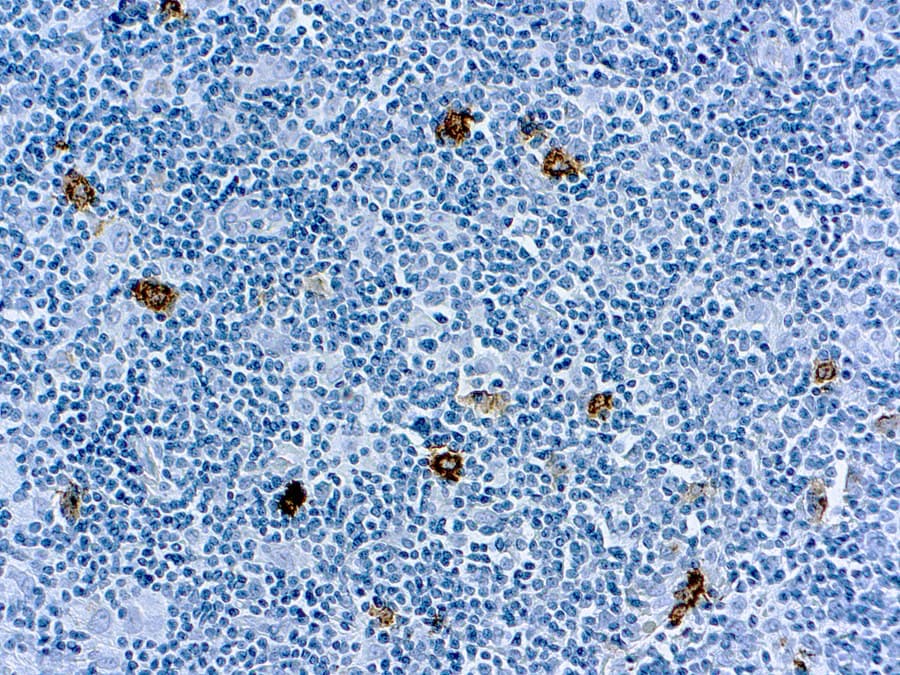
- Frequent expression in Reed–Sternberg cells of classical Hodgkin lymphoma, making CD15 an established marker in lymphoma immunophenotyping.
These properties support the use of CD15 as an immunophenotypic marker of granulocytic lineage and certain lymphoma-associated tumor cells.
Diagnostic Utility of CD15 in Hematopathology
In clinical pathology, CD15 immunohistochemistry provides valuable diagnostic information in several contexts:
- Classical Hodgkin lymphoma: Reed–Sternberg cells commonly demonstrate CD15 positivity, typically interpreted alongside CD30 and other lymphoma markers.
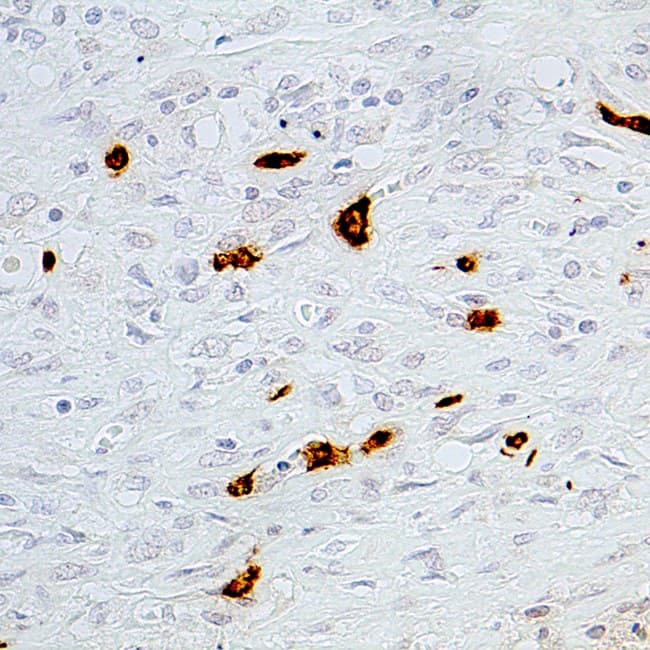
- Myeloid neoplasms: CD15 highlights maturing granulocytic cells and assists in identifying myeloid differentiation in leukemic infiltrates and myeloid sarcoma (granulocytic sarcoma).
- Leukemia immunophenotyping: CD15 expression patterns can contribute to defining immunophenotypic subsets of acute leukemias when evaluated within a broader marker panel.
Because reactive granulocytes also express CD15, interpretation requires correlation with tissue morphology and complementary immunohistochemical markers.
Key Features of CE/IVD Anti-CD15 Antibodies for IHC
CE/IVD-validated anti-CD15 antibodies are developed for standardized diagnostic immunohistochemistry workflows in clinical laboratories.
Typical features include:
- Validated performance on FFPE tissue sections used in routine pathology diagnostics.
- Specific recognition of the Lewis X epitope, enabling visualization of granulocytic lineage cells and Hodgkin lymphoma–associated tumor cells.
- Optimized staining protocols designed for reproducible immunohistochemical results.
- Compatibility with automated IHC staining platforms used in high-throughput pathology laboratories.
- Quality-controlled manufacturing, supporting consistent performance in hematopathology testing.