Anti-CD138 antibodies used in immunohistochemistry (IHC) are important diagnostic reagents for identifying plasma cells and evaluating plasma cell–associated hematologic malignancies in formalin-fixed, paraffin-embedded (FFPE) tissues.
Biological Significance of CD138 (Syndecan-1)
- Cell-surface proteoglycan: CD138 is a transmembrane heparan-sulfate proteoglycan that mediates interactions between cells and the extracellular matrix and modulates signaling pathways involving growth factors and cytokines.
- Marker of plasma cell differentiation: Within hematolymphoid tissues, CD138 is considered a reliable marker of plasmacytic differentiation and typically shows strong expression in normal and neoplastic plasma cells. However, it is not entirely plasma-cell specific because it is also expressed in epithelial tissues and various epithelial tumors.
- Role in tumor biology: Syndecan-1 contributes to the adhesion and survival of plasma cells within the bone marrow microenvironment and can influence signaling pathways involved in plasma cell proliferation and tumor–microenvironment interactions.
Diagnostic Utility of CD138 in Hematopathology
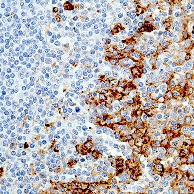
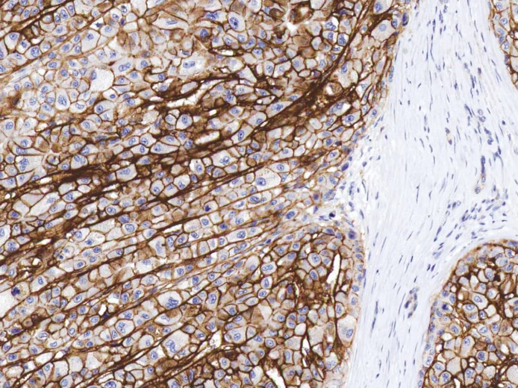
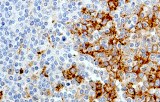
- Identification of plasma cells: CD138 typically produces strong membranous staining in plasma cells in bone marrow biopsies and lymphoid tissues, facilitating reliable visualization.
- Multiple myeloma evaluation: Most cases of multiple myeloma demonstrate strong CD138 immunoreactivity, allowing detection and quantification of neoplastic plasma cells, although rare CD138-negative plasma cell populations have been reported.
- Detection of plasmacytic differentiation: CD138 assists in identifying plasmacytic components in disorders such as lymphoplasmacytic lymphoma and other B-cell lymphomas with plasmacytic differentiation.
- Assessment of plasma cell distribution: IHC staining highlights plasma cell localization and clustering within bone marrow and tissue sections.
Key Features of Anti-CD138 CE/IVD Antibodies for IHC
- Validation for FFPE tissue sections using standard IHC workflows.
- Predominantly membranous staining consistent with syndecan-1 surface expression.
- High sensitivity for detecting plasma cells in bone marrow and lymphoid tissues.
- Compatibility with automated staining platforms and polymer-based detection systems used in clinical laboratories.
- Utility in diagnostic panels with markers such as CD38, MUM1, and immunoglobulin light-chain stains for plasma cell neoplasms.