
SOD1 antibody


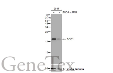
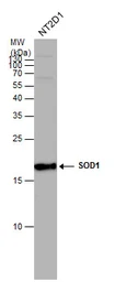
Non-transfected (–) and transfected (+) 293T whole cell extracts (30 μg) were separated by 15% SDS-PAGE, and the membrane was blotted with SOD1 antibody (GTX100554) diluted at 1:5000. The HRP-conjugated anti-rabbit IgG antibody (GTX213110-01) was used to detect the primary antibody.
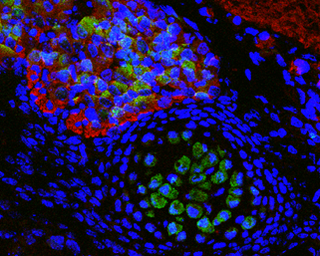
Immunofluorescence photomicrographs of paraffin-embedded mouse fetal brain.
Green: SOD1 antibody (GTX100554) diluted at 1:200. The signal was developed using goat anti-rabbit IgG antibody (Dylight488) (GTX213110-04).
Red: beta Tubulin 3/ TUJ1 antibody [GT11710] diluted at 1:100. The signal was developed using goat anti-mouse IgG antibody (Dylight594) (GTX213111-05).
Blue: Nuclear staining with Hoechst 33342.
Antigen Retrieval: Citrate buffer, pH 6.0, 15 min
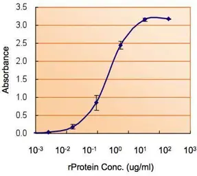
ELISA detection of SOD1 using GTX100554 for capture at a concentration of 5 μg/mL and GTX89049 for detection at a concentration of 1.5 μg/mL.
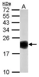
Sample (50 μg of whole cell lysate)
A: mouse brain
15% SDS PAGE
GTX100554 diluted at 1:1000
The HRP-conjugated anti-rabbit IgG antibody (GTX213110-01) was used to detect the primary antibody.
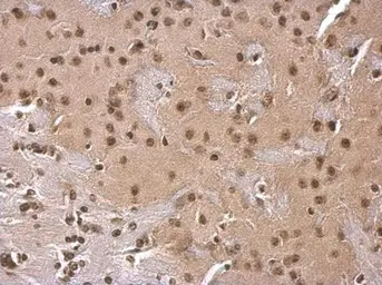
SOD1 antibody detects SOD1 protein at cytosol on mouse fore brain by immunohistochemical analysis.
Sample: Paraffin-embedded mouse fore brain.
SOD1 antibody (GTX100554) dilution: 1:500.
Antigen Retrieval: Trilogy™ (EDTA based, pH 8.0) buffer, 15min
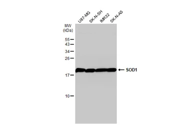
Various whole cell extracts (30 μg) were separated by 15% SDS-PAGE, and the membrane was blotted with SOD1 antibody (GTX100554) diluted at 1:1000. The HRP-conjugated anti-rabbit IgG antibody (GTX213110-01) was used to detect the primary antibody.

Sample (50 μg of whole cell lysate)
A: Rat brain
15% SDS PAGE
GTX100554 diluted at 1:1000
The HRP-conjugated anti-rabbit IgG antibody (GTX213110-01) was used to detect the primary antibody.
SOD1 antibody detects SOD1 protein by western blot analysis. Whole cell extracts (30 μg) was separated by 15% SDS-PAGE, and the membrane was blotted with SOD1 antibody (GTX100554) at a dilution of 1:1000. The HRP-conjugated anti-rabbit IgG antibody (GTX213110-01) was used to detect the primary antibody.
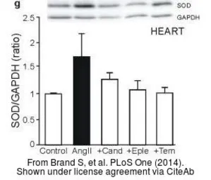
The data was published in the journal PLoS One in 2014. PMID: 25551569
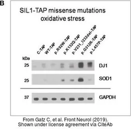
The data was published in the journal Front Neurol in 2019.PMID: 31258504
The data was published in the journal Nutrients in 2018.PMID: 30200495
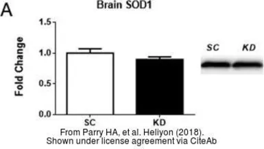
The data was published in the journal Heliyon in 2018. PMID: 30533548
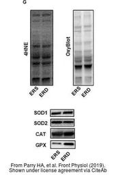
The data was published in the journal Front Physiol in 2019. PMID: 31040795
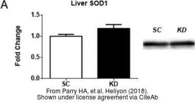
The data was published in the journal Heliyon in 2018. PMID: 30533548
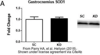
The data was published in the journal Heliyon in 2018. PMID: 30533548
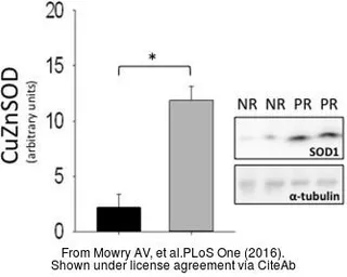
The data was published in the journal PLoS One in 2016. PMID: 27537547
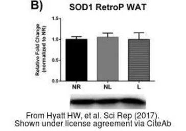
The data was published in the journal Sci Rep in 2017. PMID: 29215072
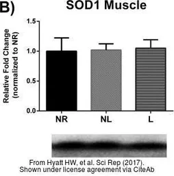
The data was published in the journal Sci Rep in 2017. PMID: 29215072
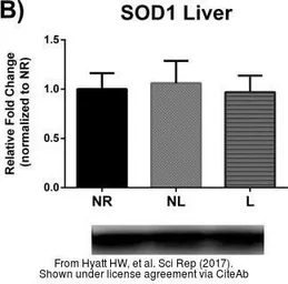
The data was published in the journal Sci Rep in 2017. PMID: 29215072
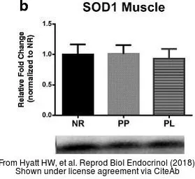
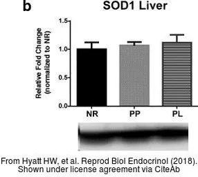
The data was published in the journal Reprod Biol Endocrinol in 2018. PMID: 29316934
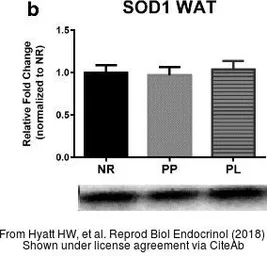
The data was published in the journal Reprod Biol Endocrinol in 2018. PMID: 29316934
The data was published in the journal Reprod Biol Endocrinol in 2018. PMID: 29316934