


Inquiry
Dispatch Time: About 3 working days
- Product Name
- FineTest®Violet 450 Anti-Human CD197/CCR7 Antibody(FN437H)
- Catalogue No.
- FV450-30090
- Form
- liquid
- Conjugation
- FineTest®Violet 450
- Conjugation Information
- FineTest® Violet 450 is designed to be excited by the violet laser (405 nm) and detected using an optical filter centered near 450 nm (e.g., a 450/45 nm bandpass filter).
- Clonality
- Monoclonal
- Isotype
- IgG2a, κ
- Clone ID
- FN437H
- Storage
- PBS with 0. 1% sodium azide, 1%BSA, pH 7.3, 2-8℃ for 12 months (Avoid repeated freeze / thaw cycles.)
Immunogen
- Alternative Names
- EBI1EVI1|CCR-7|CDw197|CMKBR7 antibody
- UniProt ID
- P32248
Application
- Tested Applications
- FC
- Recommended dilution
- Volume per test: 5μL. Each lot of this antibody is quality control tested by flow cytometric analysis. The amount of the reagent is suggested to be used 5 µL of antibody per test (million cells in 100 µL staining volume or per 100 µL of whole blood). Please check your vial before the experiment. Since applications vary, the appropriate dilutions must be determined for individual use.
Validated Images
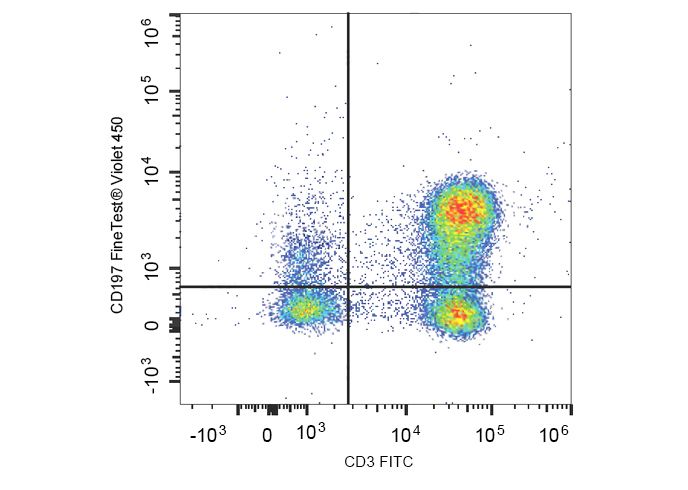 Human peripheral blood lymphocytes are stained with FineTest® Violet 450 Anti-Human CD197/CCR7 Antibody and FITC Anti-Human CD3 Antibody.
- Background
- CCR7, also known as CD197, is a chemokine receptor that binds CCL19 and CCL21. CCR7 and its ligands link innate and adaptive immunity by affecting interactions between T cells and dendritic cells and their downstream effect. Naïve T cells enter the lymph node through high endothelial venules, which express CCL21. Dendritic cells and macrophages enter the lymph node through afferent lymphatics. The encounter of T cells and dendritic cells in the T cell zone is CCR7-dependent. In addition, during immunological surveillance, B cells recirculate between B-cell-rich compartments (follicles or B cell zones) in secondary lymphoid organs, surveying for antigen. After antigen binding, B cells move to the boundary of B and T zones to interact with T-helper cells; this B cell migration is directed by CCR7 and its ligands. CCR7-positive cancer cell expression has been associated with lymph node metastasis.