Request Quote or Bulk Order
🔍
 SEC-purified HEK 293-derived EVs were stained with ExoBrite™ 410/450 CD81 Flow Antibody (left). Specific staining was seen, compared with the same antibody in buffer (right). Exosomes were detected on a CytoFLEX LX flow cytometer in the V450 (Pacific Blue) channel.  SEC-purified MCF-7-derived EVs were stained with ExoBrite™ 490/515 CD81 Flow Antibody (left). Specific staining was seen, compared with the same antibody in buffer (right). Exosomes were detected on a CytoFLEX LX flow cytometer in the B525 (FITC) channel.  SEC-purified Jurkat-derived EVs were stained with ExoBrite™ 560/585 CD81 Flow Antibody (left). Specific staining was seen, compared with the same antibody in buffer (right). Exosomes were detected on a CytoFLEX LX flow cytometer in the Y585 (PE) channel.  SEC-purified MCF-7-derived EVs were stained with ExoBrite™ R-PE CD81 Flow Antibody (left). Specific staining was seen, compared with the same antibody in buffer (right). Exosomes were detected on a CytoFLEX LX flow cytometer in the Y585 (PE) channel.  SEC-purified MCF-7-derived EVs were stained with ExoBrite™ 640/660 CD81 Flow Antibody (left). Specific staining was seen, compared with the same antibody in buffer (right). Exosomes were detected on a CytoFLEX LX flow cytometer in the R660 (APC) channel.  SEC-purified MCF-7-derived EVs were stained with ExoBrite™ APC CD81 Flow Antibody (left). Specific staining was seen, compared with the same antibody in buffer (right). Exosomes were detected on a CytoFLEX LX flow cytometer in the R660 (APC) channel.  SEC-purified MCF-7-derived EVs were stained first with ExoBrite™ 490/515 CD81 in 100 uL, followed by 1X ExoBrite™ 640/660 WGA EV Stain. EVs were detected on a CytoFLEX LX flow cytometer in the FITC and APC channels. When we gated on ExoBrite™ 640/660 WGA-positive particles, ~60% were also positive for CD81.  EVs were isolated from MCF-7 cell supernatant with CD9 affinity beads. The bead-bound EVs were stained with ExoBrite™ CD81 490/515 Flow Antibody (green) or ExoBrite™ 490/515 IgG1 Isotype Control Flow Antibody (gray), then analyzed by flow cytometry in the FITC channel.  SEC-purified mouse NIH 3T3-derived EVs were stained with ExoBrite™ 560/585 CD81 (mouse/rat) Flow Antibody (left). Specific staining was seen, compared with the same antibody in buffer (right). Exosomes were detected on a CytoFLEX LX flow cytometer in the Y585 (PE) channel.  SEC purified MCF-7-derived EVs were stained with ExoBrite™ APC CD81 Flow Antibody or a competitor CD81-APC antibody and analyzed by flow cytometry. Antibodies in buffer were used as a control to monitor aggregates. The ExoBrite™ APC antibody had more stained EVs in the gate and fewer antibody aggregates in the gate, leading to a 6-fold increase in signal-to-noise and more accurate results. EVs were detected on a CytoFLEX LX flow cytometer in the R660 (APC) channel. 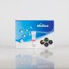 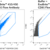 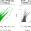 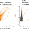 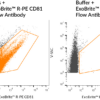 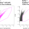 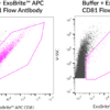 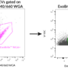 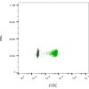 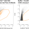 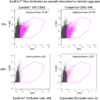
View Spectra Viewer Antibody Finder Product Description
ExoBrite™ CD81 Flow Antibody has been validated by Biotium for optimal detection of EV marker CD81 in purified or bead-bound EVs by flow cytometry. The antibody conjugates offer exceptional signal-to-noise and include color options for the Pacific Blue™, FITC, PE, and APC channels.
- Proprietary buffer formulation for enhanced signal-to-noise
- Optimal detection of EV marker CD81 by flow cytometry
- Validated for purified or bead-bound EVs
- Bright signal and low background
- Available in 6 colors
As an isotype control, we recommend ExoBrite™ IgG1 Isotype Control Flow Antibody for this antibody. Biotium also offers validated ExoBrite™ Flow Antibodies for tetraspanin proteins CD9, CD63, and other EV markers.
EV antibodies you can trust
Other commercially available antibodies against mouse and human tetraspanin proteins CD9, CD63, and CD81 are generally not validated for isolated EVs and may require tedious optimization for your EV prep and staining protocol. The antibodies and dye labels of ExoBrite™ Flow Antibody Conjugates were carefully selected and validated for robust detection of isolated EVs. In addition, the antibodies are provided in a proprietary buffer formulation for reduced antibody aggregation and brighter EV staining for optimal accuracy and signal-to-noise. Single-color and 3-color ExoBrite™ CD9/CD63/CD81 Antibody Cocktails for human tetraspanin proteins are also available for high-coverage EV staining or EV phenotyping, respectively.
For general EV staining, Biotium's ExoBrite™ True EV Membrane Stains offer unparalleled coverage of EVs in a sample and address issues of dye aggregation often seen with PKH and other common membrane dyes. Biotium also offers optimized ExoBrite™ EV Surface stains conjugated to cholera toxin B (CTB), wheat germ agglutinin (WGA), and Annexin V. These stains are specially formulated for bright and specific detection of isolated EVs by flow cytometry. These ExoBrite™ EV Surface stains may also be combined with antibody staining, for multi-parameter analysis.
Biotium offers conjugated ExoBrite™ Western Antibodies against human CD9, CD63, and CD81 designed for optimal detection in EV extracts by fluorescent western blot.
Note: In our testing, we have found that ExoBrite™ 490/515 dye may bind to streptavidin coated surfaces or beads if free biotin binding sites are not blocked. We recommend performing a biotin blocking step after binding your biotinylated capture antibody to streptavidin beads or surfaces when using ExoBrite™ 490/515 conjugates. Alternatively, consider using a different ExoBrite™ dye for staining EVs captured on streptavidin beads or surfaces.
ExoBrite™ CD81 Flow Antibody
| Antibody | Ex/Em | Target | Species Reactivity | Detection Channel | Catalog No. |
ExoBrite™ 410/450
CD81 Flow Antibody | 409/452 nm | CD81 | Human | Pacific Blue ™ | P005-410 |
ExoBrite™ 490/515
CD81 Flow Antibody | 490/516 nm | CD81 | Human | FITC | P005-490 |
ExoBrite™ 560/585
CD81 Flow Antibody | 562/584 nm | CD81 | Human | PE | P005-560 |
ExoBrite™ 640/660
CD81 Flow Antibody | 642/663 nm | CD81 | Human | APC | P005-640 |
ExoBrite™ R-PE
CD81 Flow Antibody | 496, 546, 565/578 | CD81 | Human | PE | P005-RPE |
ExoBrite™ APC
CD81 Flow Antibody | 651/660 nm | CD81 | Human | APC | P005-APC |
ExoBrite™ Flow Anti-Human CD9/CD63/CD81 Antibody Cocktails
| Cocktail | Ex/Em | Detection Channel | Size | Catalog No. |
ExoBrite™ CD9/CD63/CD81
Single-Color Antibody Cocktail (Green) | 490/516 nm | FITC | 25 tests | P030-125 |
| 100 tests | P030-500 |
ExoBrite™ CD9/CD63/CD81
Single-Color Antibody Cocktail (Orange) | 562/584 nm | PE | 25 tests | P031-125 |
| 100 tests | P031-500 |
ExoBrite™ CD9/CD63/CD81
Single-Color Antibody Cocktail (Far-Red) | 642/663 nm | APC | 25 tests | P032-125 |
| 100 tests | P032-500 |
ExoBrite™ CD9/CD63/CD81
3-Color Antibody Cocktail (Blue/Green/Far-Red) | 409/452 nm
490/516 nm
642/663 nm | Pacific Blue®
FITC
APC | 25 tests | P028-125 |
| 100 tests | P028-500 |
ExoBrite™ CD9/CD63/CD81
3-Color Antibody Cocktail (Green/Orange/Far-Red) | 490/516 nm
562/584 nm
642/663 nm | FITC
PE
APC | 25 tests | P029-125 |
| 100 tests | P029-500 |
ExoBrite™ Antibody Target Info
| Target | Relevance in EVs | References for Target in EVs | Antibody |
| CD9 | Tetraspanin; common EV marker | Kowal et al., 2016; Théry et al., 2018; Spitzberg et al., 2023 | ExoBrite™ CD9 Flow Antibody |
| ExoBrite™ CD9 (Mouse) Flow Antibody |
| ExoBrite™ CD9 Western Antibody |
| CD29 (Integrin beta 1) | Integrin cell surface glycoprotein found on EV surfaces. Has broad tissue expression and commonly found in EV proteomic analysis. | Hoshino et al., 2015; Hazawa et al., 2014; Kugeratski et al., 2021; Spitzberg et al., 2023 | ExoBrite™ CD29 Flow Antibody |
| CD47 | Associates with integrins; regulates trafficking of RNAs into EVs; may contribute to immune evasion on target cells. Has broad tissue expression and commonly found in EV proteomic analysis. | Kamerkar et al., 2017; Li et al., 2022; Kugeratski et al., 2021; Spitzberg et al., 2023 | ExoBrite™ CD47 Flow Antibody |
CD63
| Tetraspanin; common EV marker | Escola et al., 1998; Kowal et al., 2016; Spitzberg et al., 2023 | ExoBrite™ CD63 Flow Antibody |
| ExoBrite™ CD63 (Mouse) Flow Antibody |
| ExoBrite™ CD63 Western Antibody |
| CD81 | Tetraspanin; common EV marker | Escola et al., 1998; Kowal et al., 2016; Spitzberg et al., 2023 | ExoBrite™ CD81 Flow Antibody |
| ExoBrite™ CD81 (Mouse/Rat) Flow Antibody |
| ExoBrite™ CD81 Western Antibody |
| CD98 (SLC3A2) | Has broad tissue expression and commonly found in EV proteomic analysis. | Mathieu et al., 2021; Liao et al., 2024; Tissot et al., 2024; Kugeratski et al., 2021; Spitzberg et al., 2023 | ExoBrite™ CD98 Flow Antibody |
| Syntenin-1 | Involved in EV biogenesis, plays a role in tumor growth, invasion, and metastasis | Mathieu et al., 2021; Kugeratski et al., 2021; Spitzberg et al., 2023; Baietti et al., 2012 | ExoBrite™ Syntenin-1 Flow Antibody |
Technology
Extracellular Vesicle Research
Highlighted Citations
Exosome profiling with CF® dye tetraspanin panel shows single tetraspanin immunocapture methods may limit cancer biomarker detection
High-throughput fluorescence correlation spectroscopy analysis of extracellular vesicle markers
Tech Tips
Tech Tip: Extracellular Vesicle Isolation and Analysis
The Full Spectrum Blog
Bringing Light to Extracellular Vesicles
Product Attributes
Antibody type
ExoBrite™, Primary
Antibody reactivity (target)
CD81
Synonyms
CD81; Target of the antiproliferative antibody 1 (TAPA-1); Tetraspanin-28; Tspan-28
Species reactivity
Baboon, Cynomolgus monkey, Human, Mouse (low reactivity), Non-human primates
Antibody target cellular localization
Exosomes/EVs, Plasma membrane
Cell/tissue expression
Exosomes, Hepatocytes, Lymphocytes, Monocytes/macrophages
Verified antibody applications
Exosome staining (verified)
Shipping condition
Room temperature
Positive control
MCF-7 cells, MCF-7 derived exosomes
Antibody application notes
Recommended concentration for flow cytometry: 5 uL per 0.1 mL exosomes
Antibody research areas
Exosomes/EVs
Antibody/conjugate formulation
Proprietary buffer containing 0.05% sodium azide
Shelf life
Guaranteed for at least 24 months from date of receipt when stored as recommended
Storage Conditions
Store at 2 to 8 °C, Protect fluorescent conjugates from light
Regulatory status
For research use only (RUO)
Product origin
Product may contain either bovine serum albumin (BSA) from bovine serum (Bos taurus), or recombinant BSA produced in Chinese hamster ovary cells. Inquire for the specific lot.
Documents, Protocols, SDS and COA
Datasheet
Datasheet Download
COARequest the Certificate of Analysis
|