
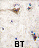
Immunohistochemistry (Formalin/PFA-fixed paraffin-embedded sections)
Formalin-fixed and paraffin-embedded human brain tissue reacted with CDH10 polyclonal antibody (Cat # PAB3148) , which was peroxidase-conjugated to the secondary antibody, followed by DAB staining. This data demonstrates the use of this antibody for immunohistochemistry; clinical relevance has not been evaluated.
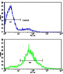
Flow Cytometry
Flow cytometric analysis of CEM cells using CDH10 polyclonal antibody (Cat # PAB3148)(bottom histogram) compared to a negative control cell (top histogram).
FITC-conjugated goat-anti-rabbit secondary antibodies were used for the analysis.

Western Blot (Cell lysate)
Western blot analysis of CDH10 polyclonal antibody (Cat # PAB3148) in CEM cell line lysates (35 ug/lane). CDH10 (arrow) was detected using the purified polyclonal antibody.
- Specifications
Product Description
Rabbit polyclonal antibody raised against synthetic peptide of CDH10.
Immunogen
A synthetic peptide (conjugated with KLH) corresponding to N-terminus of human CDH10.
Host
Rabbit
Reactivity
Human
Form
Liquid
Purification
Ammonium sulfate precipitation
Recommend Usage
Western Blot (1:1000)
Immunohistochemistry (1:10-50)
The optimal working dilution should be determined by the end user.Storage Buffer
In PBS (0.09% sodium azide)
Storage Instruction
Store at 4°C. For long term storage store at -20°C.
Aliquot to avoid repeated freezing and thawing.Note
This product contains sodium azide: a POISONOUS AND HAZARDOUS SUBSTANCE which should be handled by trained staff only.
- Applications
Immunohistochemistry (Formalin/PFA-fixed paraffin-embedded sections)
Formalin-fixed and paraffin-embedded human brain tissue reacted with CDH10 polyclonal antibody (Cat # PAB3148) , which was peroxidase-conjugated to the secondary antibody, followed by DAB staining. This data demonstrates the use of this antibody for immunohistochemistry; clinical relevance has not been evaluated.Flow Cytometry
Flow cytometric analysis of CEM cells using CDH10 polyclonal antibody (Cat # PAB3148)(bottom histogram) compared to a negative control cell (top histogram).
FITC-conjugated goat-anti-rabbit secondary antibodies were used for the analysis.Western Blot (Cell lysate)
Western blot analysis of CDH10 polyclonal antibody (Cat # PAB3148) in CEM cell line lysates (35 ug/lane). CDH10 (arrow) was detected using the purified polyclonal antibody. - Gene Info — CDH10
Entrez GeneID
1008Protein Accession#
NP_006718;Q9Y6N8Gene Name
CDH10
Gene Alias
-
Gene Description
cadherin 10, type 2 (T2-cadherin)
Omim ID
604555Gene Ontology
HyperlinkGene Summary
This gene encodes a type II classical cadherin from the cadherin superfamily, integral membrane proteins that mediate calcium-dependent cell-cell adhesion. Mature cadherin proteins are composed of a large N-terminal extracellular domain, a single membrane-spanning domain, and a small, highly conserved C-terminal cytoplasmic domain. The extracellular domain consists of 5 subdomains, each containing a cadherin motif, and appears to determine the specificity of the protein's homophilic cell adhesion activity. Type II (atypical) cadherins are defined based on their lack of a HAV cell adhesion recognition sequence specific to type I cadherins. This particular cadherin is predominantly expressed in brain and is putatively involved in synaptic adhesions, axon outgrowth and guidance. [provided by RefSeq
Other Designations
T2-cadherin|cadherin 10, type 2|cadherin-10
- Interactomes
- Diseases
- Publication Reference
- The human cadherin-10 gene: complete coding sequence, predominant expression in the brain, and mapping on chromosome 5p13-14.
Kools P, Vanhalst K, Van den Eynde E, van Roy F.
FEBS Letters 1999 Jun; 452(3):328.
- The human cadherin-10 gene: complete coding sequence, predominant expression in the brain, and mapping on chromosome 5p13-14.