

Anti-GFAP Antibody (Monoclonal, G-A-5)

Cat# MA1045
Size : 100ug/vial
Brand : Boster Bio
Contact local distributor :

Phone : +1 850 650 7790
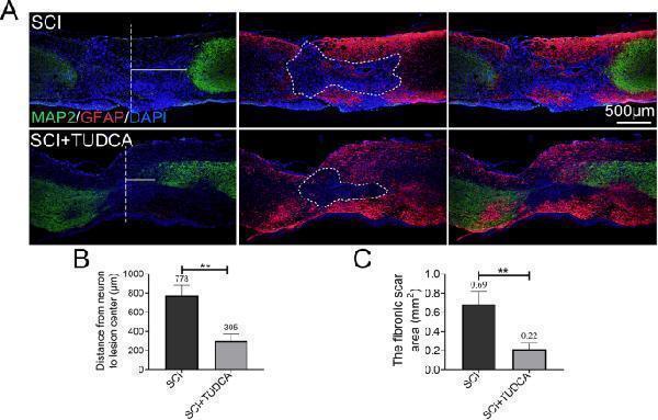
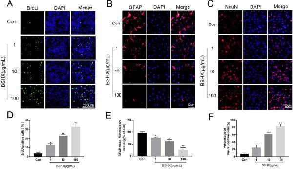
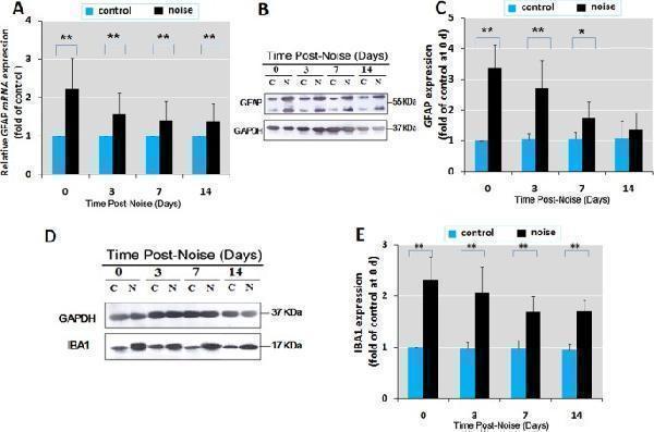
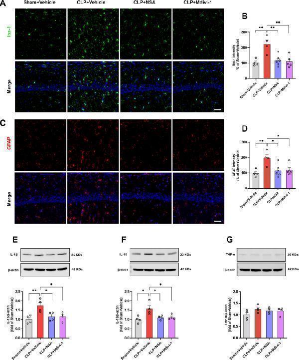
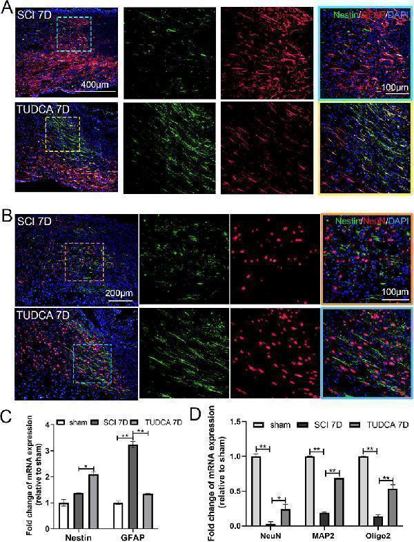
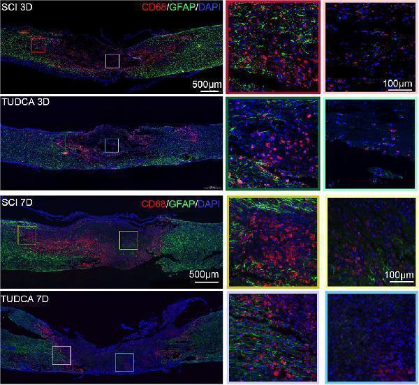
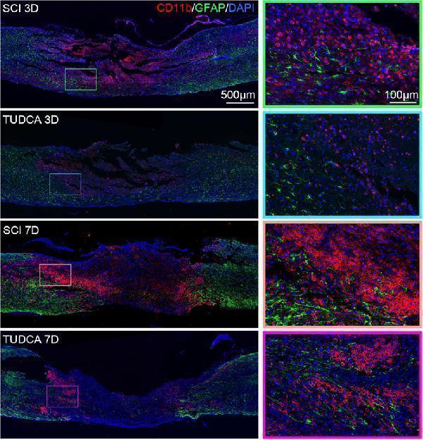
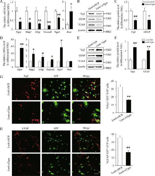
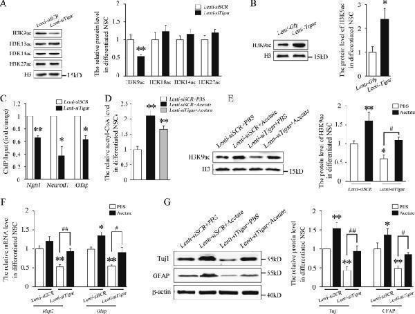
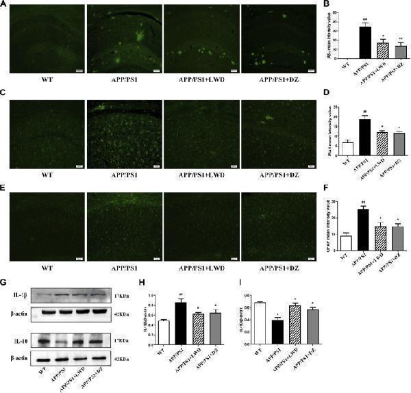
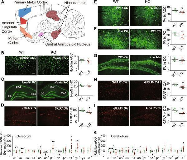
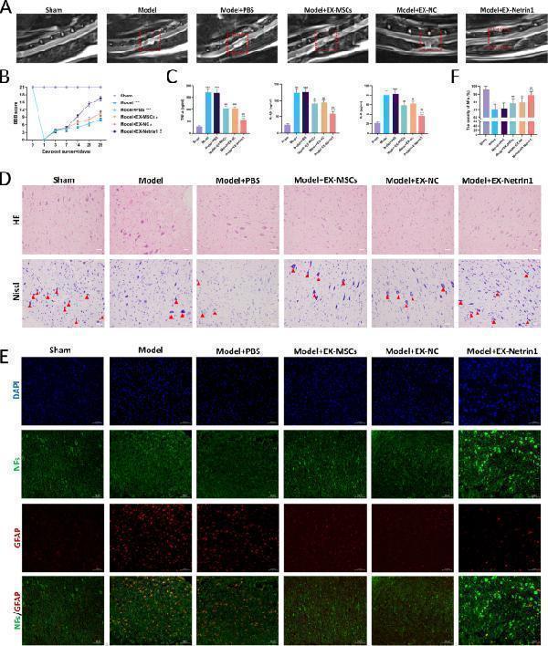
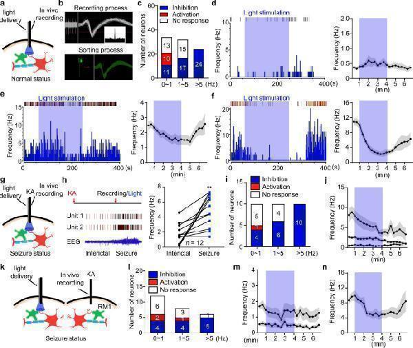
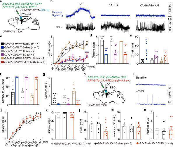
Tested Applications
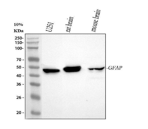
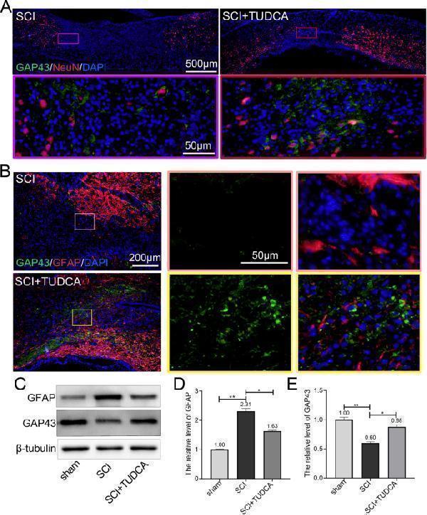
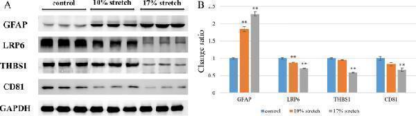
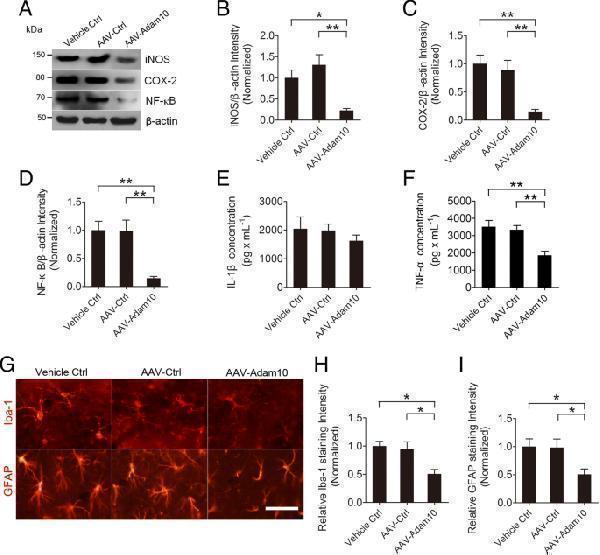
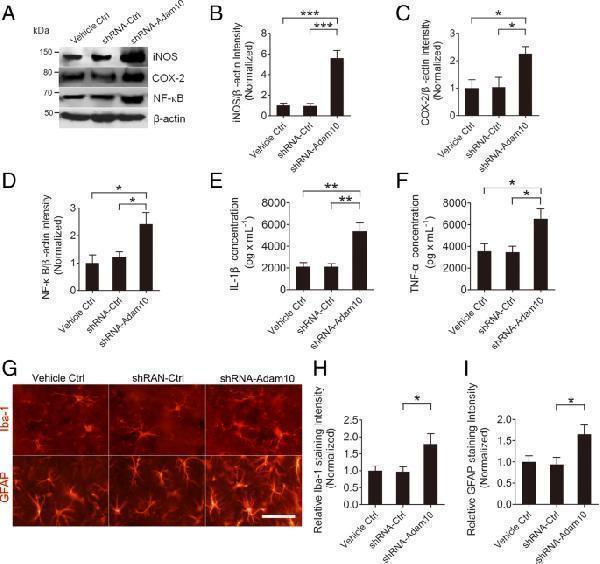
GFAP antibody was positively detected by WB in:
human U251 whole cell, rat brain tissue, mouse brain tissue
Suggested blocking solution with 5% non-fat milk or BSA; (*)Recommended protein loading: 20-40 ug per lane
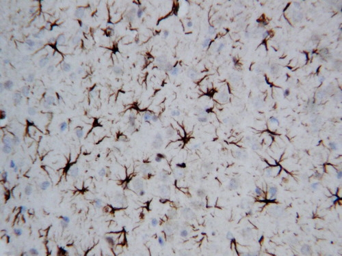
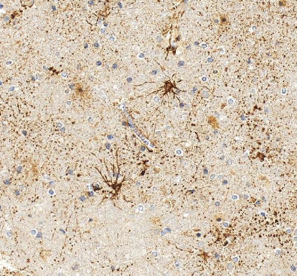
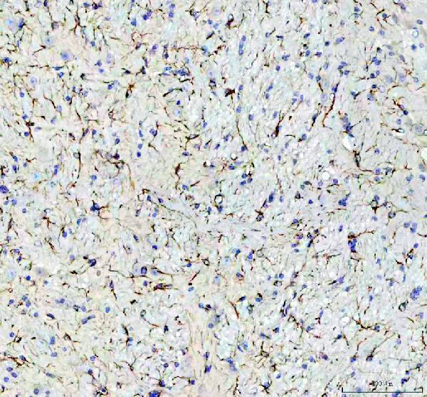
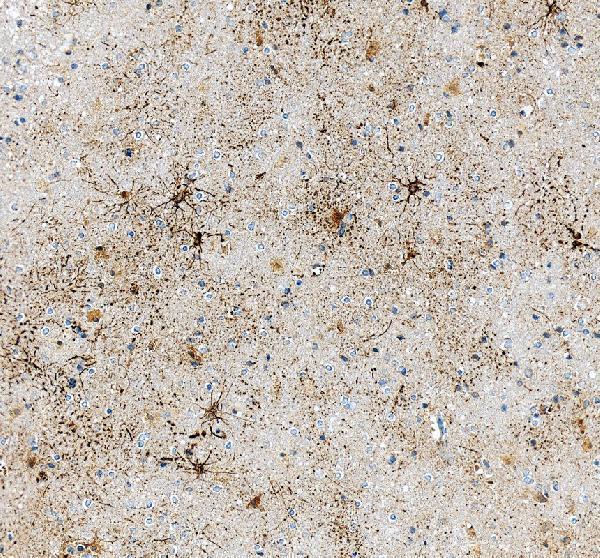
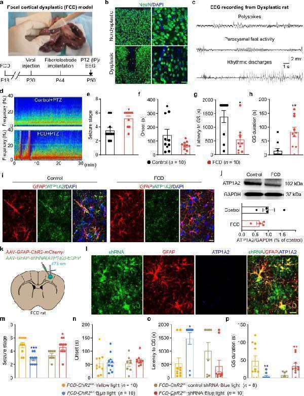
GFAP antibody was positively detected by IHC in:
rat brain tissue, mouse brain tissue, human brain tissue
Use TE buffer pH 9.0 for antigen retrieval; (*) citrate buffer pH 6.0 is an alternative.
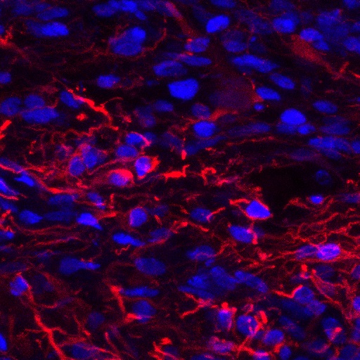
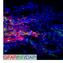
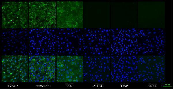
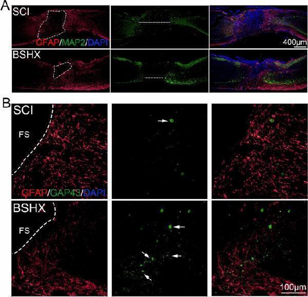
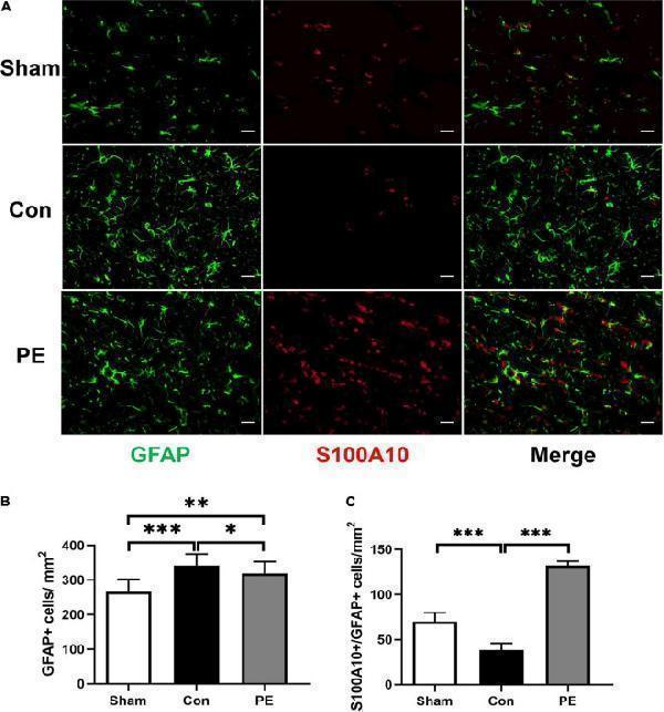
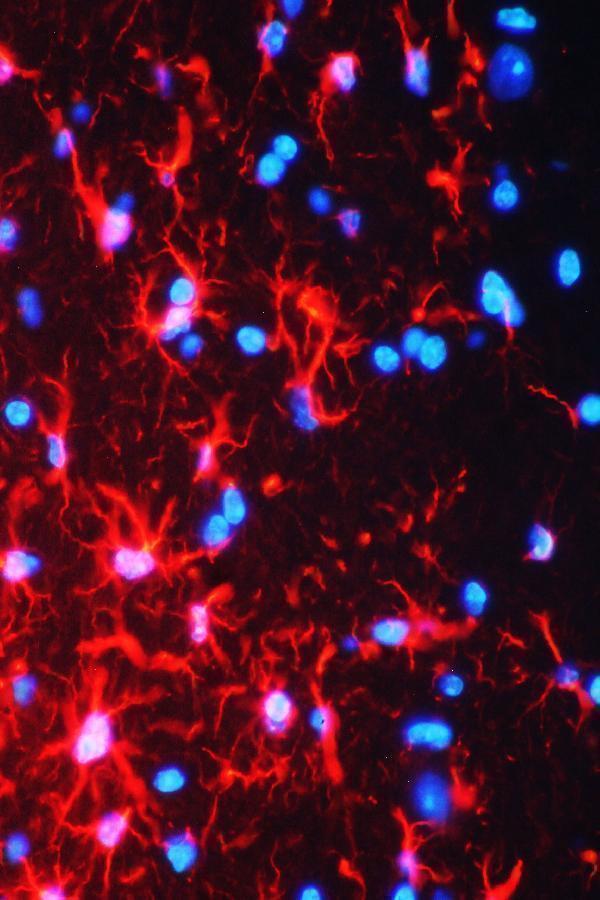
GFAP antibody was positively detected by IF in:
mouse brain tissue
Recommend Dilution
Application
Dilution
Species
Immunohistochemistry (Paraffin-embedded Section)
0.5-1μg/ml
Rat, Mouse
Western blot
0.1-0.5μg/ml
Human, Mouse, Rat
Immunofluorescence
2μg/ml
Mouse
* It is recommended to optimize the amount of this reagent in each experimental system to achieve the best results.
Product Information
Product Name
Anti-GFAP Antibody Picoband®
View all GFAP Antibodies
View all GFAP Antibodies
Form
Lyophilized
Uniprot
Gene ID
Storage & Handling
Store at -20˚C for one year from date of receipt. After reconstitution, at 4˚C for one month. It can also be aliquotted and stored frozen at -20˚C for six months. Avoid repeated freeze-thaw cycles.
Host
Rabbit
Contents
Each vial contains antibody formulated with stabilizing components, 0.9mg NaCl, 0.2mg Na2HPO4, 0.05mg Thimerosal, 0.05mg NaN3.
*This antibody is supplied in a stabilized formulation. Compatibility with conjugation reactions depends on the chemistry of the conjugation method used. For conjugation methods that are not compatible with the stabilizing components present in this formulation, a carrier-free antibody format is required.
*This antibody is supplied in a stabilized formulation. Compatibility with conjugation reactions depends on the chemistry of the conjugation method used. For conjugation methods that are not compatible with the stabilizing components present in this formulation, a carrier-free antibody format is required.
Clonality
Polyclonal
Isotype
Rabbit IgG
Immunogen
A synthetic peptide corresponding to a sequence at the C-terminus of human GFAP, identical to the related rat sequence, and different from the related mouse sequence by two amino acids.
Cross-Reactivity
No cross-reactivity with other proteins
Reconstitution
Add 0.2ml of distilled water will yield a concentration of 500ug/ml.
Observed Molecular Weight
50 kDa
Calculated Molecular Weight
49.9 kDa
Background of GFAP
Glial fibrillary acidic protein (GFAP) is an intermediate-filament (IF) protein that is highly specific for cells of astroglial lineage, although its tissue-specific role is speculative. GFAP has been located in rat kidney glomeruli and peritubular fibroblasts, leydig cells of testis, skin keratinocytes, osteocytes of bones, chondrocytes of epiglottis, bronchus, and stellate-shaped cells of the pancreas and liver. Its expression is essential for normal white matter architecture and blood-brain barrier integrity, and its absence leads to late-onset CNS dysmyelination. GFAP has also been shown to play a role in mitosis by adjusting the filament network present in the cell. During mitosis, there is an increase in the amount of phosphorylated GFAP, and a movement of this modified protein to the cleavage furrow.
Cite This Product
Anti-GFAP Antibody Picoband® (Boster Biological Technology, Pleasanton CA, USA, Catalog # PA1239)
Protocols
Western Blot protocol for GFAP antibody
Immunohistochemistry (IHC) protocol for GFAP antibody
Immunofluorescence (IF) protocol for GFAP antibody
You might also be interested by the following products:
Cat#
Description
Cond.
Price Bef. VAT