Anti-Beta-catenin antibodies validated for CE/IVD immunohistochemistry (IHC) are widely used diagnostic reagents in contemporary gastrointestinal (GI) pathology, enabling reliable evaluation of cellular adhesion status, tumor progression, and activation of the canonical Wnt signaling pathway in formalin-fixed, paraffin-embedded (FFPE) tissues. Beta-catenin, encoded by the CTNNB1 gene, is a multifunctional protein that plays a central role in both cell–cell adhesion through its interaction with E-cadherin and transcriptional regulation within the Wnt signaling cascade—two biological processes critically implicated in GI tumorigenesis, particularly in colorectal cancer and, in a subset of cases, gastric carcinoma.
In diagnostic pathology, aberrant Beta-catenin expression, most notably cytoplasmic and nuclear accumulation is a well-established surrogate marker of dysregulated Wnt signaling, frequently resulting from mutations in APC, CTNNB1, or other components of the destruction complex. Immunohistochemical detection using highly specific anti-Beta-catenin CE/IVD antibodies allows pathologists to visualize these alterations directly within the tissue context, supporting accurate diagnosis and informed clinicopathological interpretation.
Biological Significance of Beta-Catenin in Gastrointestinal Tumors
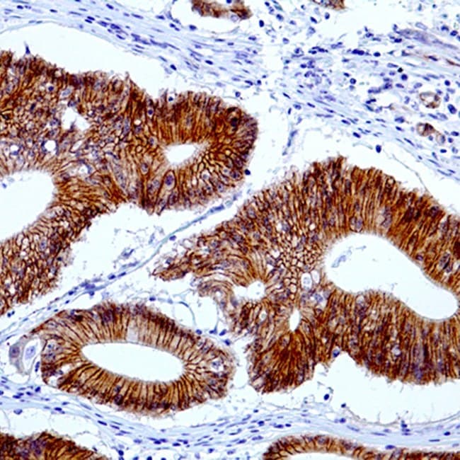
Under physiological conditions, Beta-catenin localizes predominantly to the cell membrane, where it contributes to epithelial integrity and tissue homeostasis. In GI neoplasia, disruption of the Beta-catenin destruction complex leads to protein stabilization, cytoplasmic accumulation, and nuclear translocation, resulting in transcriptional activation of Wnt target genes involved in cellular proliferation, invasion, and stemness. Extensive peer-reviewed evidence has established this shift in subcellular localization as a key molecular event in colorectal carcinogenesis and has also documented similar alterations in subsets of gastric, hepatobiliary, and intestinal tumors.
Diagnostic Utility in Gastrointestinal Pathology
Anti-Beta-catenin IHC antibodies play an important role in routine diagnostic practice and in advanced molecular pathology applications within GI pathology.
Key Diagnostic Applications
Colorectal adenomas and adenocarcinomas
- A progressive shift from membranous to cytoplasmic and nuclear staining correlates with increasing degrees of dysplasia and malignant transformation.
- Supports the identification of Wnt-driven colorectal carcinoma subtypes in conjunction with histomorphology and molecular findings.
Differential diagnosis of GI and intra-abdominal spindle cell lesions
- Diffuse nuclear Beta-catenin expression supports a diagnosis of desmoid-type fibromatosis, although it is not entirely specific.
- Useful in distinguishing desmoid-type fibromatosis from gastrointestinal stromal tumors (GISTs; typically, negative), inflammatory myofibroblastic tumors, and most other mesenchymal neoplasms.
- Rare nuclear positivity has been reported in histologic mimics such as solitary fibrous tumor and should be interpreted within the appropriate clinicopathological context.
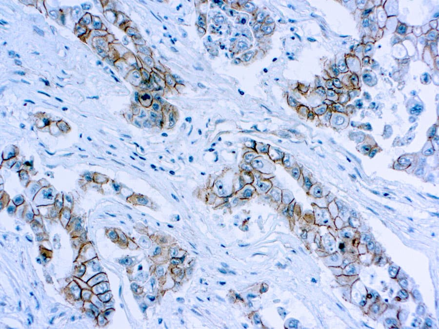
Gastric carcinoma and other GI malignancies
- Aberrant Beta-catenin expression patterns have been reported in subsets of gastric carcinoma and other GI cancers and have been associated with invasive behavior, tumor progression, and lymph node involvement.
- May contribute to prognostic assessment when interpreted alongside established clinicopathological parameters and complementary immunomarkers.
Key Features of Anti-Beta-Catenin CE/IVD Antibodies
- High specificity and sensitivity for Beta-catenin detection in FFPE gastrointestinal tissues.
- Reliable visualization of membranous, cytoplasmic, and nuclear staining patterns critical for diagnostic interpretation.
- Optimized performance on automated IHC staining platforms commonly used in diagnostic pathology laboratories.
- CE/IVD validation ensuring regulatory compliance, analytical robustness, and inter-laboratory reproducibility.