Anti-ALK antibodies for IHC can enable sensitive detection of tumor-associated ALK protein expression in FFPE tissue. When assays are analytically/clinically validated and interpreted with morphology, lineage markers, and—when indicated—molecular studies, ALK IHC supports confident diagnosis and classification of ALK-rearranged hematolymphoid neoplasms.
Biological significance of ALK
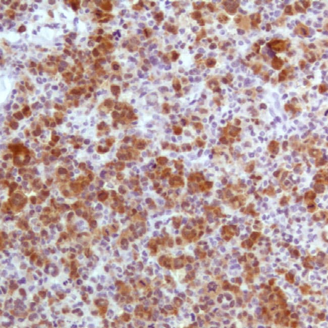
ALK is a receptor tyrosine kinase that is oncogenic in hematologic malignancies predominantly through ALK gene fusions (e.g., NPM1::ALK) that drive constitutive signaling. In systemic ALK+ ALCL, ALK fusions define a biologically distinct entity, and the subcellular IHC pattern often reflects the fusion partner (e.g., nuclear + cytoplasmic staining in many NPM1::ALK cases).
Diagnostic utility of ALK IHC in hematopathology
- Entity confirmation & differential diagnosis: ALK IHC helps confirm systemic ALK+ ALCL and supports distinction from mimics when integrated with morphology and lineage markers.
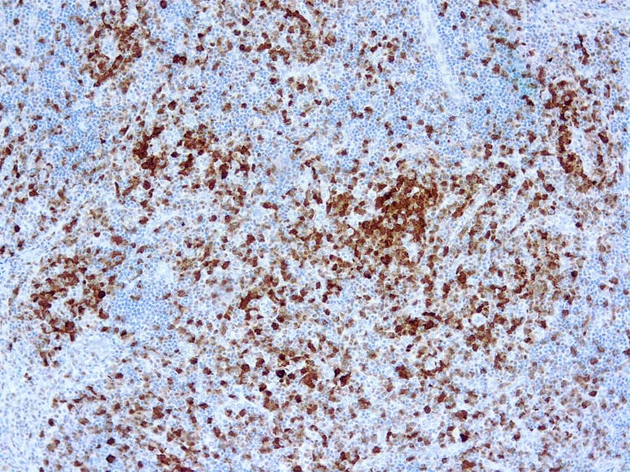
- Disease mapping: Bone marrow involvement in ALK+ ALCL may be difficult to detect morphologically and can be highlighted by ALK immunostaining in appropriate contexts.
- Rare ALK+ lymphomas: In ALK+ LBCL, ALK IHC is pivotal; staining is often cytoplasmic, and perinuclear/Golgi-zone patterns have been described with certain fusion partners.
Key features reported for high-performance ALK IHC antibodies
- Validated clones & pattern fidelity: A 2023 study found a 5A4-based protocol non-inferior to ALK01 for diagnosing ALK+ ALCL.
- Interpretive localization: Fusion-partner–dependent localization (nuclear/cytoplasmic patterns) can add interpretive value beyond a binary positive/negative read.