Anti-Lambda (λ) CE/IVD antibodies for immunohistochemistry (IHC) are widely used adjunct tools in hematopathology for the evaluation of B-cell clonality and plasma cell disorders. Immunoglobulin light chain analysis (κ/λ) is routinely incorporated as part of the diagnostic workup to help distinguish reactive from neoplastic lymphoid proliferations, particularly in formalin-fixed, paraffin-embedded (FFPE) tissues.
Biological Significance of Lambda (λ) Light Chains
Immunoglobulins consist of two heavy chains and two light chains, with λ representing one of the two light chain isotypes expressed by B cells. Each B cell expresses a single light chain type (κ or λ), reflecting allelic exclusion during B-cell development and immunoglobulin gene rearrangement. Clonal λ light chain expression is associated with immunoglobulin lambda (IGL) gene rearrangements and may indicate a monoclonal B-cell population.
Diagnostic Utility in Hematopathology
- Demonstration of monotypic λ expression supports the presence of a clonal B-cell or plasma cell population and is used as part of the diagnostic assessment of lymphoid and plasma cell neoplasms.
- Reactive lymphoid tissues typically show a polytypic κ/λ distribution, whereas neoplastic proliferations often demonstrate light chain restriction.
- Light chain analysis provides supportive diagnostic information and is interpreted in conjunction with histomorphology and additional immunophenotypic or molecular findings.
- λ restriction can be observed in plasma cell neoplasms, including multiple myeloma and related plasma cell disorders, although κ-restricted cases are also common depending on disease subtype.
Key Features of Anti-Lambda CE/IVD Antibodies (IHC)
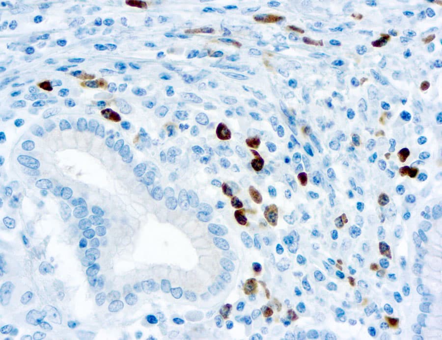
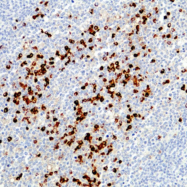
- High specificity for λ light chains expressed in B lymphocytes and plasma cells, enabling detection of both surface and cytoplasmic immunoglobulin.
- Validated for use in FFPE tissues, supporting routine application in surgical pathology laboratories.
- Strong reactivity in plasma cells, facilitating identification of monoclonal populations in appropriate diagnostic contexts.
- Compatibility with automated IHC staining platforms and standardized laboratory workflows in accordance with CE/IVD requirements.
- Supportive role in differential diagnosis, including evaluation of suspected B-cell lymphomas, plasmacytomas, and atypical lymphoid infiltrates.