Anti-KBA.62 (Melanoma Associated Antigen) CE/IVD antibodies are immunohistochemistry reagents developed for the detection of a melanoma-associated cell membrane antigen expressed in a broad range of benign and malignant melanocytic proliferations. Originally generated against a human melanoma cell line, KBA.62 recognizes an incompletely characterized melanoma-associated antigen that is distinct from conventional melanocytic differentiation markers such as HMB-45 and Melan-A/MART-1. Unlike melanogenesis-associated markers, KBA.62 expression is frequently preserved in poorly differentiated and amelanotic melanomas, making it particularly valuable in diagnostically challenging cases.
Biological Significance of Melanoma Associated Antigen (KBA.62)
- Recognizes an incompletely characterized melanoma-associated cell membrane antigen distinct from conventional melanocytic differentiation antigens.
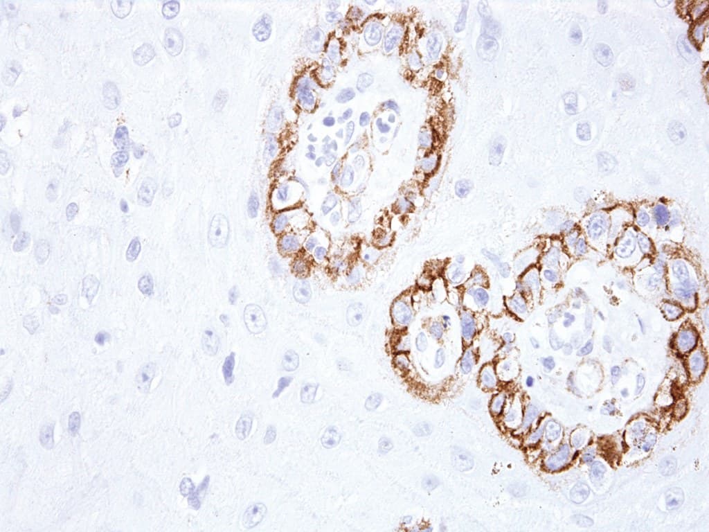
- Demonstrates predominantly membranous immunoreactivity, differing from the predominantly cytoplasmic staining pattern observed with HMB-45 and Melan-A/MART-1.
- Expressed in benign melanocytic nevi and in a high proportion of primary and metastatic melanomas, including spindle cell and desmoplastic variants.
- May remain expressed in melanomas showing loss of conventional melanocytic differentiation markers.
Diagnostic Utility in Dermatopathology
- Supports the diagnosis of cutaneous melanoma, particularly in poorly differentiated or diagnostically challenging lesions.
- Useful for identifying amelanotic melanomas that may show reduced or absent expression of HMB-45 or Melan-A/MART-1.
- Demonstrates diagnostic utility in desmoplastic melanoma, spindle cell melanoma, and metastatic melanoma.
- Frequently incorporated into melanoma immunopanels together with S100, SOX10, PRAME, Melan-A, and HMB-45 to improve diagnostic sensitivity.
- May assist in the evaluation of sentinel lymph node metastases and metastatic melanoma deposits.
- As KBA.62 expression may occur in selected non-melanocytic neoplasms, results should be interpreted in conjunction with histomorphology and complementary immunohistochemical markers.
Key Features of CE/IVD Anti-KBA.62 Antibodies
- Optimized for immunohistochemical detection in formalin-fixed, paraffin-embedded (FFPE) tissues.
- Designed for CE/IVD diagnostic use in clinical pathology laboratories.
- Produces predominantly membranous staining with excellent morphological contrast.
- Compatible with major automated IHC staining platforms according to manufacturer validation and instructions for use (IFU).
- Suitable for dermatopathology and oncologic pathology workflows requiring sensitive detection of melanocytic lesions and melanoma.