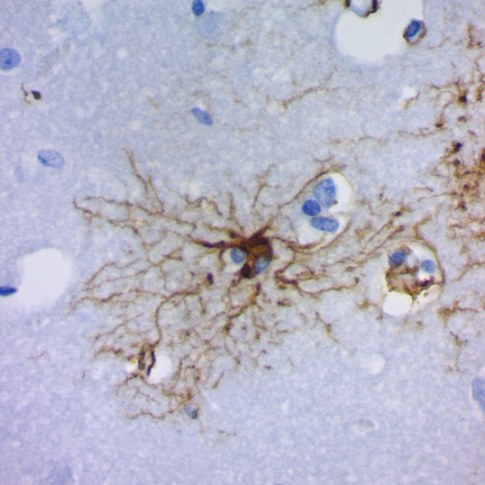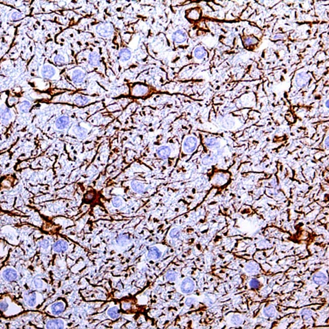
Glial Fibrillary Acidic Protein (GFAP) is a type III intermediate filament protein predominantly expressed in astrocytes of the central nervous system. It is widely recognized in peer-reviewed neuropathology literature as a key structural component of the astroglial cytoskeleton, contributing to cell shape maintenance, mechanical stability, and astrocyte reactivity during central nervous system injury and disease. In addition to mature astrocytes, GFAP expression may also be observed in certain neural progenitor populations during development, as reported in the neuropathology literature.
In neuropathology, GFAP is extensively used as a robust astrocytic marker in immunohistochemistry (IHC), enabling the identification and characterization of reactive gliosis and supporting the assessment of astrocytic lineage differentiation in central nervous system tumors. Its expression is frequently upregulated in response to CNS injury, neuroinflammation, and neurodegenerative processes, making it a widely used biomarker for both diagnostic and research applications. However, GFAP expression may be variable in some high-grade or dedifferentiated glial neoplasms, and therefore it is generally interpreted in conjunction with other diagnostic markers and histopathological findings.
Biological Significance of GFAP
- Major intermediate filament protein of astrocytes in the central nervous system.
- Contributes to astrocyte structural integrity, cytoskeletal organization, and glial scar formation.
- Upregulated during reactive astrogliosis in CNS injury and neurodegenerative disease.
- Established marker of astrocytic lineage in developmental and adult brain studies.
Diagnostic Utility of GFAP in Neuropathology
- Widely used immunohistochemical marker for astrocytic differentiation in central nervous system tumors (e.g., astrocytomas, glioblastomas), with recognized variability in expression depending on tumor grade and cellular differentiation status.
- Used to detect and evaluate reactive gliosis in CNS injury and inflammatory conditions.
- Supports differential diagnosis of glial versus non-glial neoplasms in brain and spinal cord biopsies when interpreted alongside complementary markers.
- Widely implemented in neuro-oncology and surgical neuropathology workflows as part of broader immunohistochemical panels.
Key Features of Anti-GFAP CE/IVD Antibodies for IHC
- CE/IVD status indicates conformity with applicable in vitro diagnostic regulatory frameworks, depending on manufacturer and jurisdictional approval.
- High analytical sensitivity and specificity for GFAP detection in formalin-fixed, paraffin-embedded (FFPE) tissue.
- Optimized for immunohistochemistry (IHC) with validated protocols supporting reproducible staining in diagnostic workflows.
- Batch-to-batch consistency designed to support standardized performance in neuropathology laboratories.
- Compatible with manual and automated staining platforms in clinical histopathology settings.