CD99 (MIC2) is a heavily glycosylated 32 kDa type I transmembrane glycoprotein encoded by the MIC2 gene located within the pseudoautosomal region 1 (PAR1) of both the X and Y chromosomes. The protein is involved in multiple biological processes, including cell adhesion, transendothelial migration, apoptosis, cellular differentiation, intracellular trafficking, and immune cell signaling. Physiologically, CD99 is expressed in cortical thymocytes, subsets of hematopoietic progenitor cells, endothelial cells, and selected mesenchymal tissues, accounting for its important role in developmental biology and pediatric pathology.
Diagnostic Utility of CD99 in Pediatric Pathology
Anti-CD99 antibodies are indispensable components of immunohistochemical panels used for the evaluation of pediatric small round blue cell tumors and poorly differentiated neoplasms.
Key diagnostic applications include:
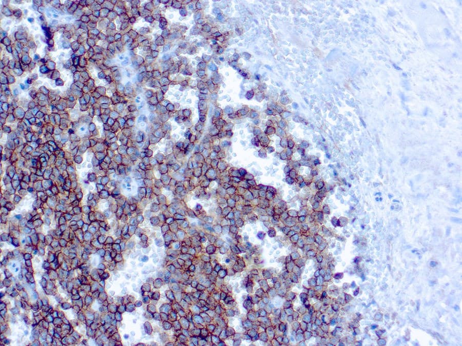
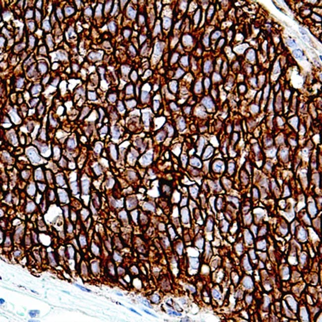
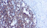
- Ewing sarcoma: typically demonstrates strong, diffuse membranous CD99 expression in approximately 90–95% of cases, with some series reporting positivity approaching 100%.
- Differential diagnosis of pediatric round cell tumors, including:
- lymphoblastic lymphoma/leukemia,
- CIC-rearranged sarcoma,
- mesenchymal chondrosarcoma,
- desmoplastic small round cell tumor,
- rhabdomyosarcoma,
- small cell osteosarcoma.
- Correlation with molecular testing, particularly the detection of EWSR1-ETS gene rearrangements in suspected Ewing sarcoma.
Although highly sensitive for Ewing sarcoma, CD99 expression is not specific and may be observed in several other pediatric neoplasms. Consequently, interpretation should always be performed in conjunction with morphology, additional immunohistochemical markers, and molecular findings.
Key Features of CE/IVD Anti-CD99 Antibodies
- Optimized for immunohistochemical staining of formalin-fixed, paraffin-embedded (FFPE) tissue sections.
- Produce the characteristic crisp membranous staining pattern required for diagnostic interpretation in Ewing sarcoma.
- Validated for routine diagnostic IHC workflows on both manual and automated staining platforms.
- High analytical sensitivity combined with excellent preservation of tissue morphology and architectural context.
- Suitable for incorporation into multiparametric pediatric sarcoma panels including NKX2.2, FLI1, ERG, WT1, desmin, myogenin, and cytokeratins.