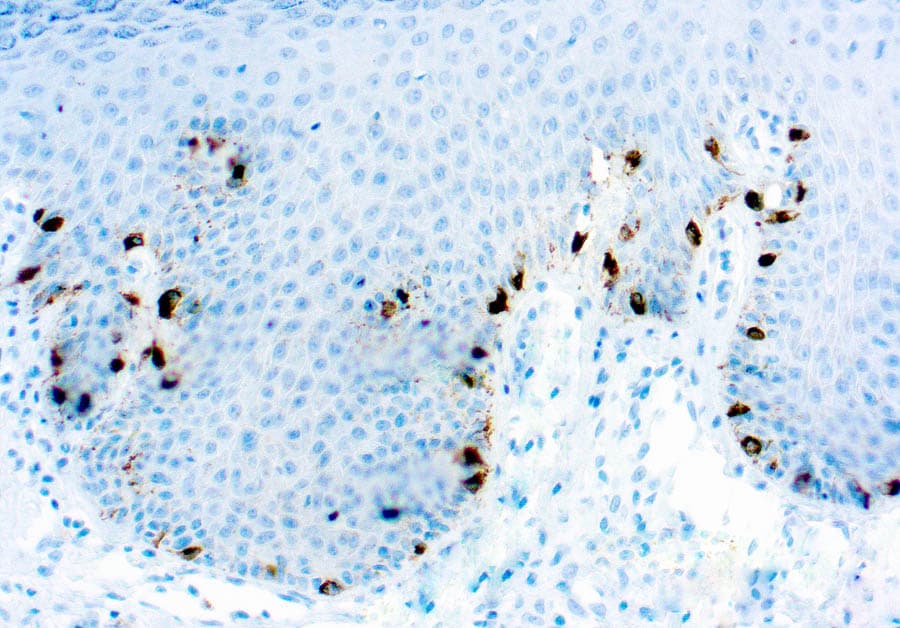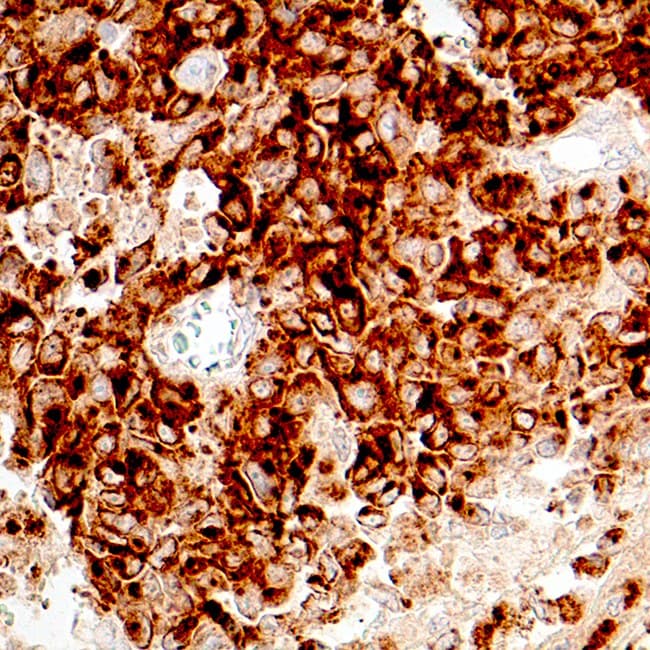
Tyrosinase (TYR) is a melanosomal copper-containing oxidase that catalyzes key rate-limiting reactions in melanogenesis, including the hydroxylation of L-tyrosine to L-DOPA and the subsequent oxidation of L-DOPA to dopaquinone. As a critical enzyme in melanin biosynthesis, TYR plays a central role in melanocyte differentiation and pigment production. TYR expression is highly restricted to normal melanocytes and melanocytic neoplasms, making it a valuable melanocytic lineage-associated biomarker in dermatopathology and melanoma diagnostics.
Biological Significance of Tyrosinase
- Essential enzyme involved in melanin biosynthesis and pigment production.
- Expressed predominantly in normal melanocytes and melanocytic tumors.
- Transcriptionally regulated by melanocytic differentiation pathways involving MITF and other melanogenesis-associated proteins.
- Localized primarily to melanosomes, resulting in characteristic cytoplasmic granular expression in melanocytic cells.
Diagnostic Utility of Tyrosinase in Dermatopathology
- Supports the identification of melanocytic differentiation in benign and malignant melanocytic proliferations.
- Useful in the diagnostic evaluation of both primary and metastatic melanoma.
- Commonly incorporated into melanocytic immunohistochemical panels alongside S100, SOX10, Melan-A/MART-1, and HMB-45 to improve diagnostic confidence and classification accuracy.
- Demonstrates high specificity for melanocytic differentiation, although sensitivity may be lower than SOX10 or S100 in spindle cell and desmoplastic melanoma, where expression can be reduced or absent.
Key Features of CE/IVD Anti-Tyrosinase Antibodies
- Validated for use on formalin-fixed paraffin-embedded (FFPE) tissue specimens.
- Optimized for routine immunohistochemistry workflows, including compatibility with automated staining platforms.
- Typically produce distinct granular cytoplasmic staining corresponding to melanosomal localization.
- Selected CE/IVD antibody clones are optimized to provide high analytical specificity and minimal background staining under validated assay conditions.
- Developed and manufactured in accordance with applicable CE-marked IVD regulatory requirements for clinical diagnostic laboratories.