Podoplanin (PDPN) is a heavily O-glycosylated mucin-type transmembrane sialoglycoprotein with an apparent molecular weight of approximately 36–43 kDa, depending on its degree of glycosylation. It is physiologically expressed in lymphatic endothelial cells, mesothelial cells, podocytes, type I pneumocytes, follicular dendritic cells, and selected stromal cell populations. Functionally, podoplanin participates in lymphangiogenesis, tissue development, platelet activation through interaction with CLEC-2, and regulation of cell migration and cytoskeletal remodeling. In neoplasia, PDPN expression has been implicated in tumor invasion, epithelial–mesenchymal transition-associated phenotypes, and metastatic dissemination.
Biological Significance of Podoplanin
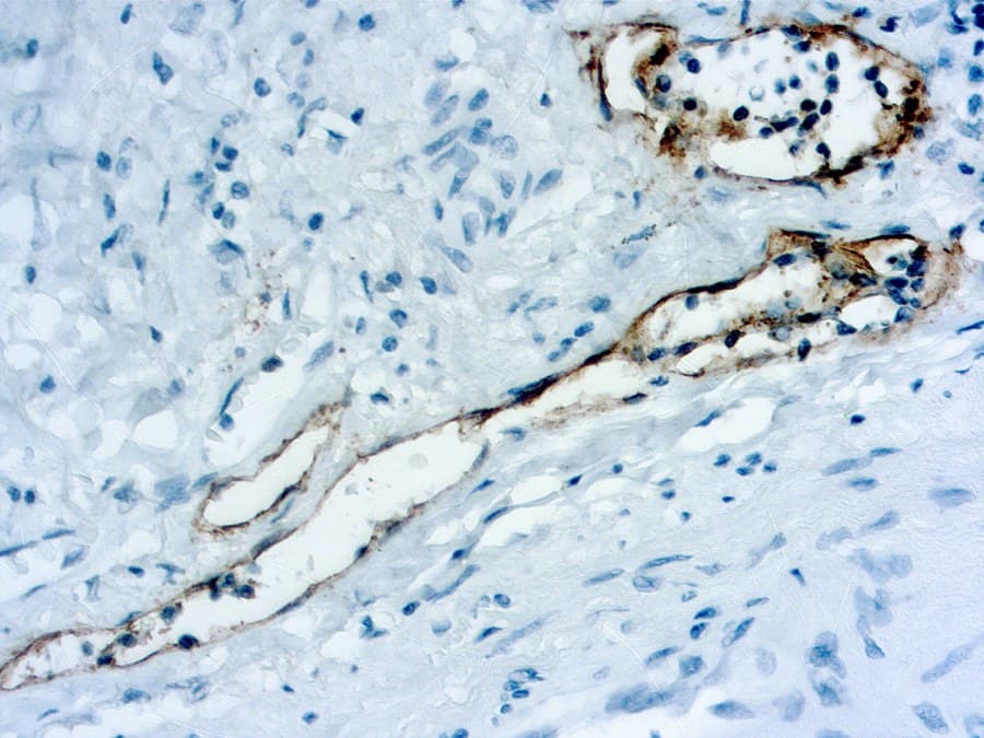
- Established marker of lymphatic endothelial differentiation, with expression generally absent from blood vascular endothelium.
- Plays an essential role in lymphatic vessel development, maturation, and maintenance.
- Mediates platelet activation, aggregation, and tumor–stromal interactions through CLEC-2 signaling.
- Contributes to cell motility, migration, and invasive behavior in multiple tumor types.
Diagnostic Utility of Podoplanin in Soft Tissue Pathology
- Widely used for the identification of lymphatic vessels and the assessment of lymphovascular invasion in soft tissue tumors.
- Supports the diagnosis of tumors showing lymphatic differentiation, including lymphangioma, kaposiform hemangioendothelioma, and angiosarcomas exhibiting lymphatic differentiation.
- Commonly expressed in Kaposi sarcoma, where it may assist in distinguishing lymphatic from blood vascular neoplasms when interpreted alongside endothelial markers such as CD31, CD34, and ERG.
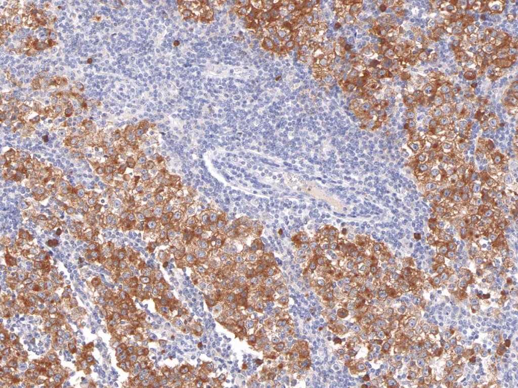
- Demonstrates immunoreactivity in selected spindle cell and mesenchymal neoplasms, including some peripheral nerve sheath tumors and dendritic cell tumors; therefore, interpretation requires correlation with morphology and a broader immunohistochemical panel.
- Although highly valuable for the assessment of lymphatic lineage, podoplanin lacks sufficient specificity to serve as a stand-alone marker in the differential diagnosis of most soft tissue sarcomas.
Key Features of CE/IVD Anti-Podoplanin Antibodies
- Validated for use on formalin-fixed paraffin-embedded (FFPE) tissues.
- Typically supplied as mouse monoclonal antibodies, most commonly using the D2-40 clone.
- Produce a characteristic membranous staining pattern in positive cells.
- Optimized for both automated and manual immunohistochemistry workflows.
- Manufactured according to CE/IVD quality standards for routine diagnostic pathology laboratories.