MART-1 (Melan-A) is a melanocyte lineage-specific differentiation antigen encoded by the MLANA gene and expressed predominantly in normal melanocytes, melanocytic nevi, and most conventional melanomas. The protein participates in early melanosome biogenesis by regulating the stability, trafficking, and processing of proteins involved in melanosome formation and maturation, particularly PMEL and GPR143/OA1. Although initially identified as a melanoma-associated antigen recognized by cytotoxic T lymphocytes, MART-1 is now established as one of the principal markers of melanocytic differentiation in surgical pathology and dermatopathology.
Biological Significance of MART-1 (Melan-A)
- Melanocyte lineage-specific differentiation antigen with highly restricted tissue expression.
- Localized predominantly to the endoplasmic reticulum, Golgi apparatus, and early melanosomal compartments.
- Contributes to melanosome biogenesis, maturation, and melanogenesis.
- Exhibits relatively homogeneous expression in most conventional melanocytic lesions and many melanoma subtypes.
Diagnostic Utility in Dermatopathology
- Identification and characterization of benign and malignant melanocytic proliferations.
- Confirmation of melanocytic differentiation in primary and metastatic melanoma.
- Assessment of junctional melanocyte distribution and pagetoid spread in melanocytic lesions.
- Evaluation of melanoma margins, including frozen sections and Mohs micrographic surgery specimens.
- Commonly incorporated into immunohistochemical panels with SOX10, S100, and HMB-45 to improve diagnostic sensitivity and specificity.
- Reduced sensitivity may occur in desmoplastic melanoma, spindle-cell melanoma, and dedifferentiated metastatic melanoma.
Key Features of MART-1 (Melan-A) CE/IVD Antibodies
- Validated for immunohistochemical staining of formalin-fixed, paraffin-embedded (FFPE) tissues.
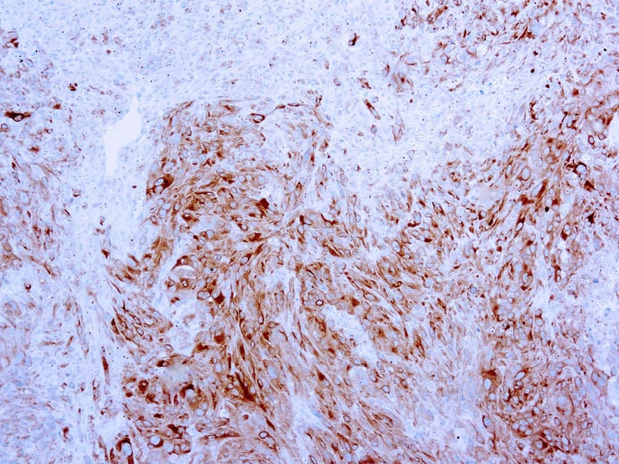
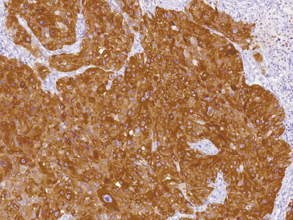
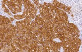
- Typically produce strong, granular cytoplasmic staining of melanocytic cells with excellent visualization of melanocyte morphology.
- Widely available in optimized ready-to-use (RTU) and concentrated formulations suitable for manual and automated staining platforms.
- Most CE/IVD assays employ the extensively validated A103 mouse monoclonal antibody clone (IgG1 isotype).
- High specificity for melanocytic differentiation, although expression may occasionally be observed in steroid-producing tissues and selected non-melanocytic neoplasms.