CD1a (Cluster of Differentiation 1a) is a human MHC class I-like glycoprotein that presents lipid and glycolipid antigens to subsets of T cells and is highly expressed on Langerhans cells and related dendritic cells. This molecule serves as an essential immunohistochemical marker in diagnosing hematopathologic entities such as Langerhans cell histiocytosis (LCH).
Biological Significance of CD1a
- CD1a is part of the CD1 family of antigen-presenting molecules that bind lipid antigens and display them to T cell receptors, enabling lipid-specific T cell activation distinct from classical peptide MHC pathways.
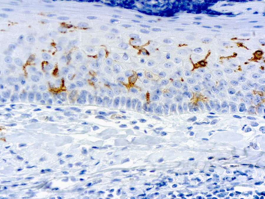
- It is constitutively expressed at high levels on epidermal Langerhans cells and certain dendritic cell subsets and interacts with lipid antigens in the secretory pathway for T cell engagement.
Diagnostic Utility in Hematopathology
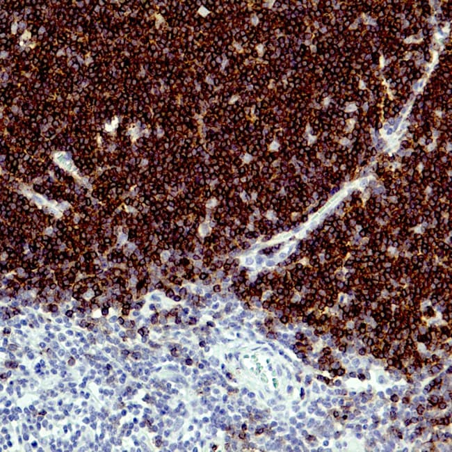
- In Langerhans cell histiocytosis (LCH), strong membranous CD1a immunoreactivity in lesional cells is a defining diagnostic marker when interpreted with morphology and other markers (e.g., CD207/Langerin, S100).
- Presence of CD1a helps distinguish LCH from other histiocytic and dendritic neoplasms where CD1a is absent or low.
- CD1a IHC can also characterize cortical thymocyte lineage in specific T cell lymphoproliferative disorders, although expression varies by neoplasm.
Key Features of Anti-CD1a CE/IVD Antibodies for IHC
- High specificity and sensitivity: Validated monoclonal clones for CE-marked IVD use bind human CD1a with minimal cross-reactivity, enabling clear membranous staining patterns in FFPE tissue.
- Clinical diagnostic performance: These antibodies facilitate reproducible detection of CD1a positive cells, aiding classification in complex hematopathology panels when used with other lineage markers.
- Optimized for automated platforms: CE/IVD reagents are formulated for standardized antigen retrieval and compatibility with automated immunostainers, supporting quality control in diagnostic laboratories.