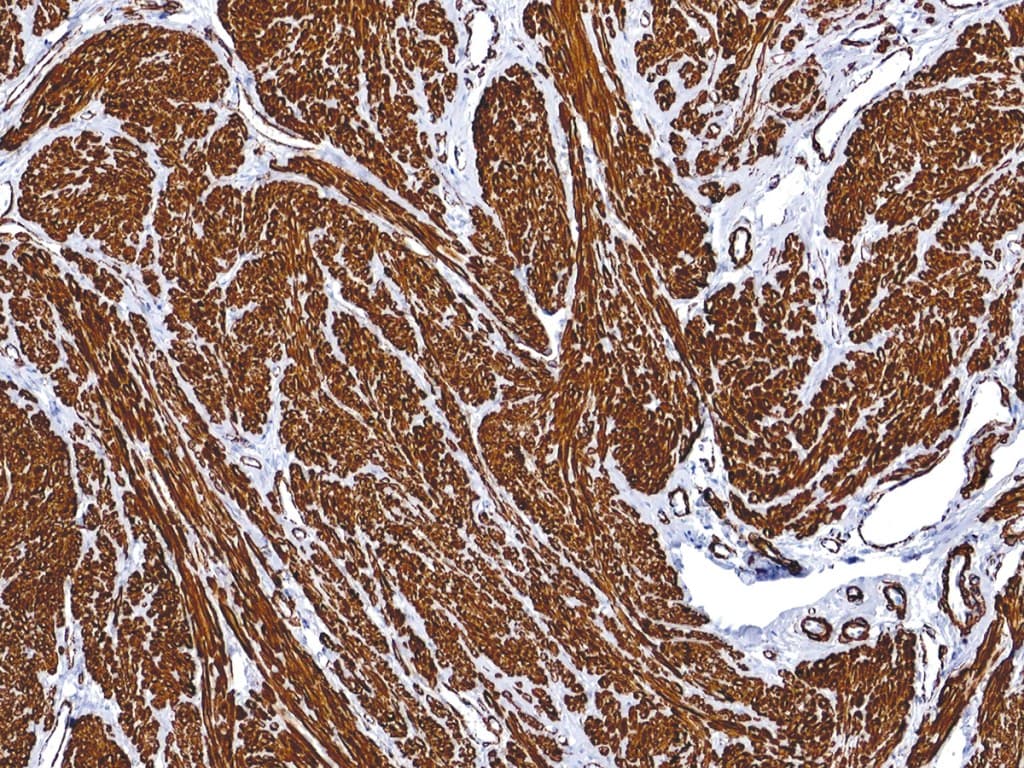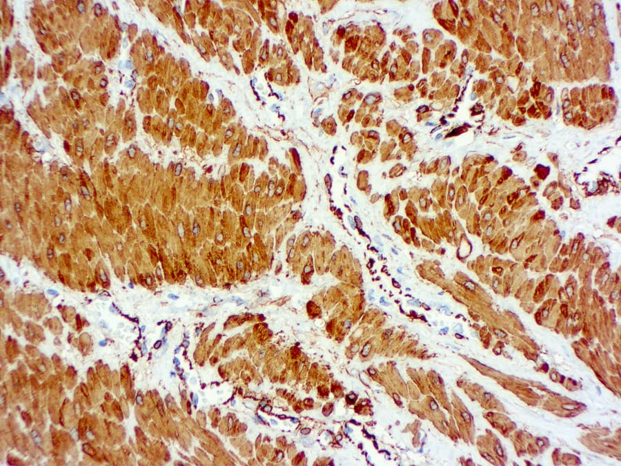
Alpha Smooth Muscle Actin (α-SMA), encoded by the ACTA2 gene, is a 42 kDa actin isoform that constitutes a major component of the thin filaments of the contractile apparatus in vascular and visceral smooth muscle cells. In addition to its role in contractility, α-SMA contributes to cytoskeletal organization, mechanotransduction, cellular migration, extracellular matrix remodeling, and tissue repair through its expression in activated myofibroblasts. In normal tissues, α-SMA expression is predominantly observed in smooth muscle cells, pericytes, myofibroblasts, and myoepithelial cells, making it a sensitive marker of smooth muscle and myofibroblastic differentiation in diagnostic pathology.
Biological Significance of α-SMA
- Major component of the thin filaments of the smooth muscle contractile apparatus.
- Regulates cellular contractility through interactions with myosin motor proteins.
- Marker of activated myofibroblasts involved in wound healing, fibrosis, and tissue remodeling.
- Participates in mechanosensing, cellular migration, and extracellular matrix deposition.
Diagnostic Utility of α-SMA in Soft Tissue Pathology
α-SMA immunohistochemistry is routinely employed for the identification and classification of neoplasms exhibiting smooth muscle or myofibroblastic differentiation. It is particularly useful in the evaluation of:
- Leiomyoma and leiomyosarcoma.
- Myofibroblastic neoplasms, including inflammatory myofibroblastic tumor and myofibroma.
- Perivascular tumors such as glomus tumors and myopericytic neoplasms.
- Spindle cell lesions when interpreted in conjunction with desmin, h-caldesmon, calponin, and muscle-specific actin.
- Stromal myofibroblast activation and tumor microenvironment remodeling.
Because α-SMA expression is not restricted to smooth muscle cells, it is considered a sensitive but relatively non-specific marker and is typically interpreted as part of a broader immunohistochemical panel.
Key Features of CE/IVD α-SMA Antibodies
- Validated for formalin-fixed paraffin-embedded (FFPE) tissues.
- Optimized for standardized and reproducible IHC workflows.
- Commonly available as highly specific monoclonal antibodies, including the widely used clone 1A4.
- Characteristic cytoplasmic staining pattern with minimal background reactivity under optimized conditions.
- Compatible with automated staining platforms and routine diagnostic laboratories.
- Designed to support CE/IVD-compliant clinical pathology applications requiring high analytical reproducibility and manufacturing consistency.