

Anti-Cytochrome C/CYCS Antibody Picoband®

Cat# PA1118
Size : 100ug/vial
Brand : Boster Bio
Contact local distributor :

Phone : +1 850 650 7790
Tested Applications
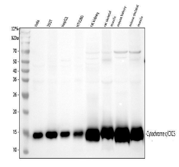
CYCS antibody was positively detected by WB in:
human Hela whole cell, human 293T whole cell, human HepG2 whole cell, human HT1080 whole cell, rat kidney tissue, rat skeletal muscle tissue, mouse kidney tissue, mouse skeletal muscle tissue
Suggested blocking solution with 5% non-fat milk or BSA; (*)Recommended protein loading: 20-40 ug per lane
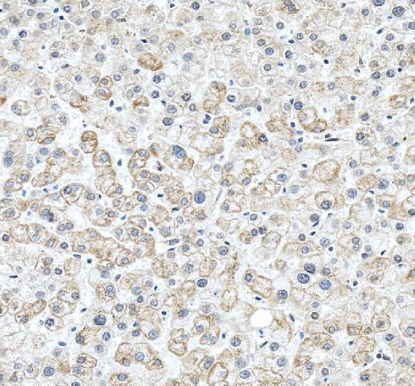
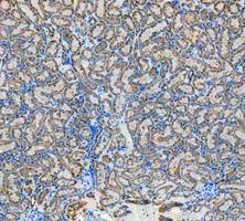
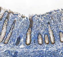
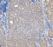
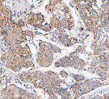
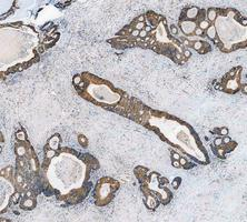
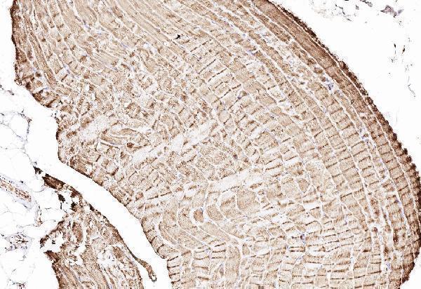
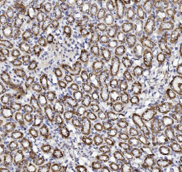
CYCS antibody was positively detected by IHC in:
human skeletal muscle tissue, rat kidney tissue, human liver tissue, rat kidney tissue, human appendix tissue, human liver tissue, human colon tissue, human cervial tissue
Use TE buffer pH 9.0 for antigen retrieval; (*) citrate buffer pH 6.0 is an alternative.
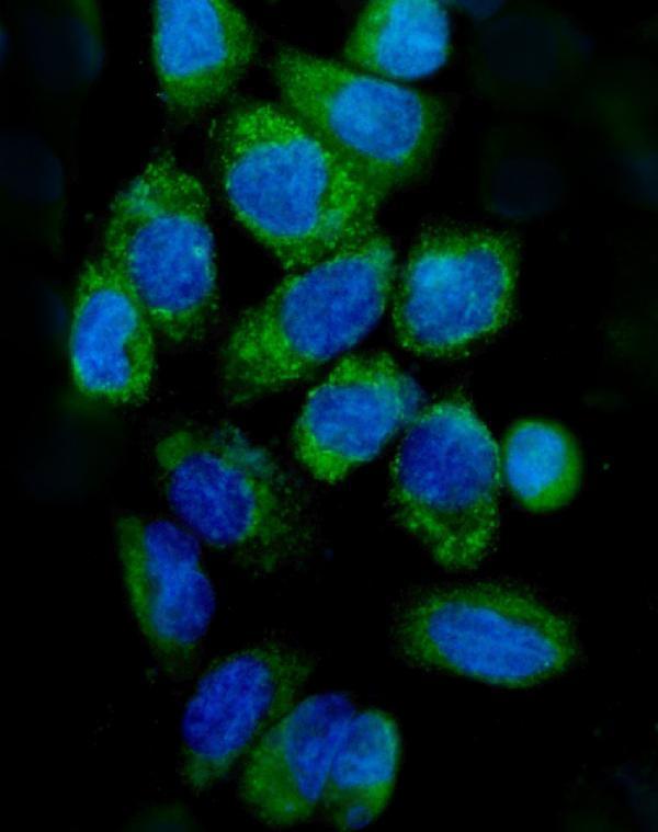
CYCS antibody was positively detected by ICC/IF in:
A431 cell
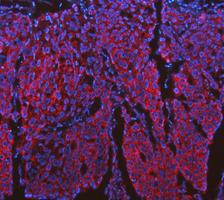
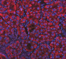
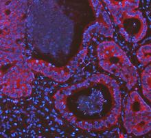
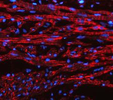
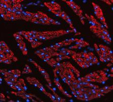
CYCS antibody was positively detected by IF in:
human cervical cancer tissue, human liver cancer tissue, human colon cancer tissue, rat heart tissue, mouse heart tissue
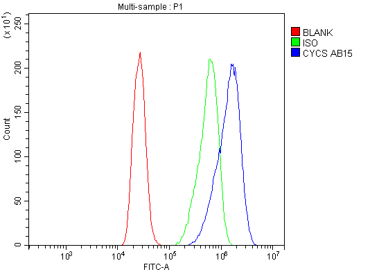
CYCS antibody was positively detected by FC in:
CACO-2 cell
Recommend Dilution
* It is recommended to optimize the amount of this reagent in each experimental system to achieve the best results.
Note: For flow cytometry (FCM), validated for fixed cells only. Not validated for FACS or surface flow cytometry using live cells.
Product Information
View all CYCS Antibodies
*This antibody is supplied in a stabilized formulation. Compatibility with conjugation reactions depends on the chemistry of the conjugation method used. For conjugation methods that are not compatible with the stabilizing components present in this formulation, a carrier-free antibody format is required.
11.7 kDa