Human Bcl-xL Antibody : Biotin

Referência OASB01179
Tamanho : 0.1mg
Marca : Aviva Systems Biology
Contactar o distribuidor local :
Human Bcl-xL Antibody : Biotin (OASB01179)
| Datasheets/Manuals | Printable datasheet for Human Bcl-xL Antibody : Biotin (OASB01179) |
|---|
| Tested Species Reactivity | Human |
|---|---|
| Predicted Species Reactivity | Mouse, Rat, Rhesus |
| Clonality | Monoclonal |
| Clone | 7B2.5 |
| Isotype | IgG3 |
| Host | Mouse |
| Conjugation | Biotin |
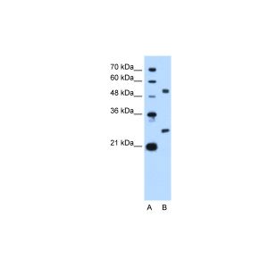
| Application | FC, IHC-P, ICC, WB, IP |
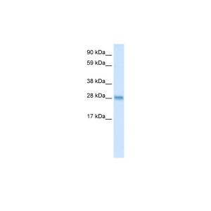
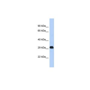
| Additional Information | Description: Bcl-xL is a 29 kDa member of the Bcl-2 family of proteins involved in regulation of programmed cell death, or apoptosis. It is found in both soluble and membrane fractions of cellular lysates. Like Bcl-2, Bcl-xL has been demonstrated to block apoptosis which is induced by a variety of stimuli and, under certain conditions, offers greater protection against apoptosis than Bcl-2. While formation of heterodimers with the apoptosis-enhancing protein, Bax does not block the apoptosis-suppressing property of Bcl-xL, heterodimerization with Bad effectively inhibits the protective function of Bcl-xL. |
| Reconstitution and Storage | Store at 2-8C |
| Immunogen | Recombinant human Bcl-xS |
| Concentration | 0.1 mg/mL |
| Specificity | Bcl-xL |
| Characterization | To insure lot- to- lot consistency, each batch of product is tested by immunoassay to conform to thecharacteristics of a standard reference reagent. |
| Warning | Reagents contain sodium azide. Sodium azide is very toxic if ingested or inhaled. Avoid contact with skin,eyes, or clothing. Wear eye or face protection when handling. If skin or eye contact occurs, wash withcopious amounts of water. If ingested or inhaled, contact a physician immediately. Sodium azide yieldstoxic hydrazoic acid under acidic conditions. Dilute azide- containing compounds in running water beforediscarding to avoid accumulation of potentially explosive deposits in lead or copper plumbing. |
| Dilution | Flow Cytometry: Purified antibody Fluorescein conjugate Biotin conjugate R-Phycoerythrin conjugate <= 3 ug/106 cells <= 3 ug/106 cells <= 3 ug/106 cells <= 0.3 ug/106 cells Western Blotting: Purified antibody <= 0.5 ug/ml |
| Application Info | Immunoprecipitation15,16, Immunohistochemistry17, Flow cytometry17, Western blotting18 |
| Other Applications Data | Since applications vary, you should determine the optimum workingdilution of the product that is appropriate for your specific need. |
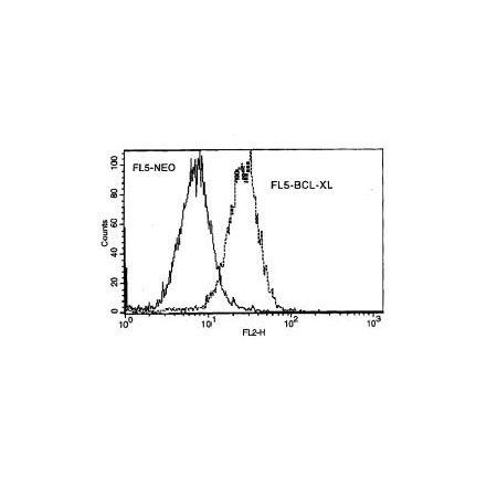
| Other Applications Image 1 Data | Amount Used: 2 Ug/106 cells Murine FL5 cells (FL5-NEO) and FL5 cells transfected with Bcl-xL expression plasmid (FL5-BCL-XL) were fixed with buffered paraformaldehyde and then permeabilized with saponin. The cells were incubated with mouse anti-h |
| Storage | - The purified (UNLB) antibody is supplied as 0.1 mg of purified immunoglobulin in 1.0 mL of 100 mMborate buffered saline, pH 8.2. No preservatives or amine- containing buffer salts added. Store at 2- 8 C - The fluorescein (FITC) conjugate is supplied as 0.1 mg in 1.0 mL of PBS/NaN3. Store at 2- 8 C - The biotin (BIOT) conjugate is supplied as 0.1 mg in 1.0 mL of PBS/NaN3. Store at 2- 8 C - The R- phycoerythrin (R- PE) conjugate is supplied as 0.1 mg in 1.0 mL of PBS/NaN3 and a stabilizingagent. Store at 2- 8 C. Do not freeze! - Protect conjugated forms from light. Reagents are stable for the period shown on the label if stored asdirected. |
| Reference | 1. Cohen, J.J. 1991. Adv. Immunol. 50:55 2. Cohen, J.J., and R.C. Duke. 1992. Annu. Rev. Immunol. 10:267 3. Nunez, G., and M.F. Clarke. 1994. Trends Cell Biol. 4:399 4. Cory, S. 1995. Annu. Rev. Immunol. 13:513 5. Oltvai., Z.N., C.L. Millman, and S.J. Korsmeyer. 1993. Cell 74:609 6. Farrow, S.N., et al. 1995. Nature 374:731 7. Chittenden, T., et al. 1995. Nature 374:733 8. Kiefer, M.C., et al. 1995. Nature 374:736 9. Boise, L.H., M. Gonzalez- Garcia, C.E. Postema, L. Ding, T. Lindstein, L.A. Turka, X. Mao, G. Nunez, and C.B. Thompson 1993. Cell 74:597 10. Gottschalk, A.R., L.H. Boise, C.B. Thompson, and J. Quintans. 1994. Proc. Natl. Acad. Sci. USA 91:7350 11. Gonzalez- Garcia, M., I. Garcia, L. Ding, S. O'Shea, L.H. Boise, C.B. Thompson, and G. Nunez. 1995. Proc. Natl. Acad. Sci. USA 92:430412. Dole, M.G., R. Jasty, M.J. Cooper, C.B. Thompson, G. Nunez, and V.P. Castle. 1995. Can. Res. 55:2576 13. Shimizu, S., et al. 1995. Nature 374:811 14. Yin, X.M., Z.N. Oltvai, and S.J. Korsmeyer. 1994. Nature 369:321 15. Cheng, E.H.- Y., et al. 1996. Nature 379:554 16. Gottschalk, A.R., L.H. Boise, Z.N. Oltvai, M.A. Accavitti, S.J. Korsmeyer, J. Quintans, and C.B. Thompson. 1996. Cell Death and Differentiation 3:113 17. Boise, L.H. 1997. Personal communication 18. L. Haughan. 1997. Personal communication. |
|---|---|
| Gene Symbol | BCL2L1 |
| Gene Full Name | Bcl-2-like protein 1 |
| Alias Symbols | BCLX, BCL2L, Bcl-X, PPP1R52, BCL-XL/S |
| NCBI Gene Id | 598; 12048; 24888; 713035 |
| Protein Name | bcl-2-like protein 1 |
| Description of Target | The protein encoded by this gene belongs to the BCL-2 protein family. BCL-2 family members form hetero- or homodimers and act as anti- or pro-apoptotic regulators that are involved in a wide variety of cellular activities. The proteins encoded by this gene are located at the outer mitochondrial membrane, and have been shown to regulate outer mitochondrial membrane channel (VDAC) opening. VDAC regulates mitochondrial membrane potential, and thus controls the production of reactive oxygen species and release of cytochrome C by mitochondria, both of which are the potent inducers of cell apoptosis. Alternative splicing results in multiple transcript variants encoding two different isoforms. The longer isoform acts as an apoptotic inhibitor and the shorter isoform acts as an apoptotic activator. |
| Uniprot ID | Q07817, Q64373, P53563, F6UKR4 |
| Protein Accession # | NP_001182.1 |
| Nucleotide Accession # | NM_001191.3 |